Copyright
©2011 Baishideng Publishing Group Co.
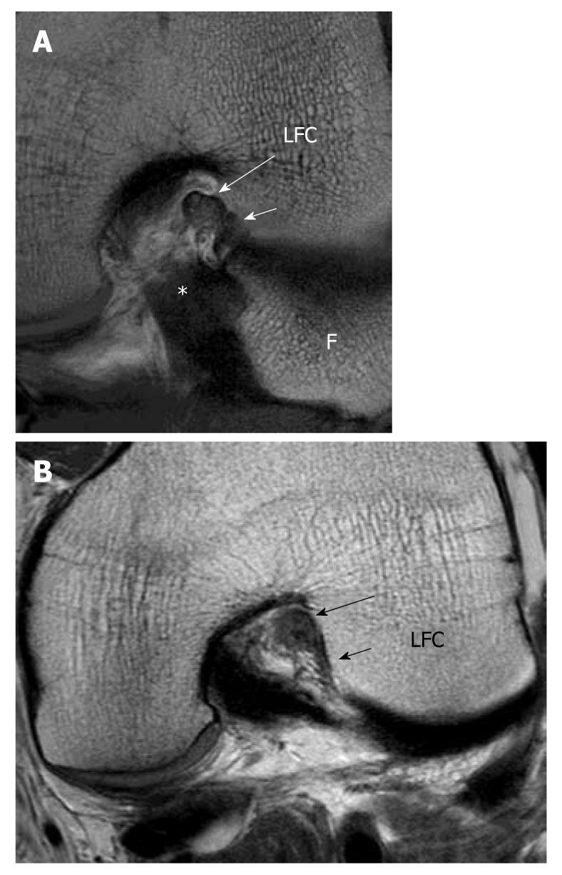
Figure 7 High resolution imaging anterior cruciate ligament in oblique axial plane.
A: Oblique axial image can clearly delineate the two bundles and assess each bundle separately; AM bundle (long white arrow) PL bundle (short white arrow). B: Partial tear of the anterior cruciate ligament. Oblique axial image at the femoral side shows thickening and hyperintense signal intensity of the AM bundle (black long arrow) while fibres are absent in the region of the PL bundle (black short arrow). Features are compatible with high grade partial AM bundle tear and complete PL tear which were confirmed in arthroscopy. LFC: Lateral femoral condyle; *: Posterior cruciate ligament; F: Fibular head.
- Citation: Ng WHA, Griffith JF, Hung EHY, Paunipagar B, Law BKY, Yung PSH. Imaging of the anterior cruciate ligament. World J Orthop 2011; 2(8): 75-84
- URL: https://www.wjgnet.com/2218-5836/full/v2/i8/75.htm
- DOI: https://dx.doi.org/10.5312/wjo.v2.i8.75


