Copyright
©2011 Baishideng Publishing Group Co.
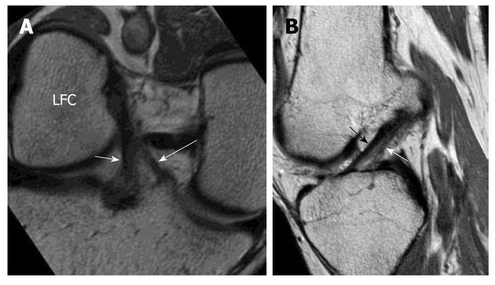
Figure 6 High resolution imaging normal anterior cruciate ligament in oblique coronal and oblique sagittal planes.
Volunteer of a 31-year-old man with no history of injury and clinical instability. Note that the AM bundle (white long arrow) and PL bundle (white short arrow) can be well depicted on the oblique coronal image at the tibial attachment but not at the femoral attachment and not by the oblique sagittal image.
- Citation: Ng WHA, Griffith JF, Hung EHY, Paunipagar B, Law BKY, Yung PSH. Imaging of the anterior cruciate ligament. World J Orthop 2011; 2(8): 75-84
- URL: https://www.wjgnet.com/2218-5836/full/v2/i8/75.htm
- DOI: https://dx.doi.org/10.5312/wjo.v2.i8.75


