Copyright
©2011 Baishideng Publishing Group Co.
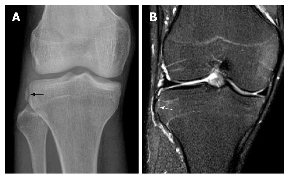
Figure 2 Segond fracture.
A 30-year-old man suffered knee injury during a basketball match. A: Frontal radiograph of the right knee shows an avulsion fracture fragment at the lateral tibial rim (black arrow) compatible with Segond fracture. This fracture is frequently associated with a torn anterior cruciate ligament which also happened in this patient (not shown); B: A coronal T2-weighted fat suppression magnetic resonance image of the same patient. Corresponding site reveals a minimally displaced Segond fracture (white arrow) which may be easily missed in this patient with no significant bone bruise or oedema.
- Citation: Ng WHA, Griffith JF, Hung EHY, Paunipagar B, Law BKY, Yung PSH. Imaging of the anterior cruciate ligament. World J Orthop 2011; 2(8): 75-84
- URL: https://www.wjgnet.com/2218-5836/full/v2/i8/75.htm
- DOI: https://dx.doi.org/10.5312/wjo.v2.i8.75


