Copyright
©2011 Baishideng Publishing Group Co.
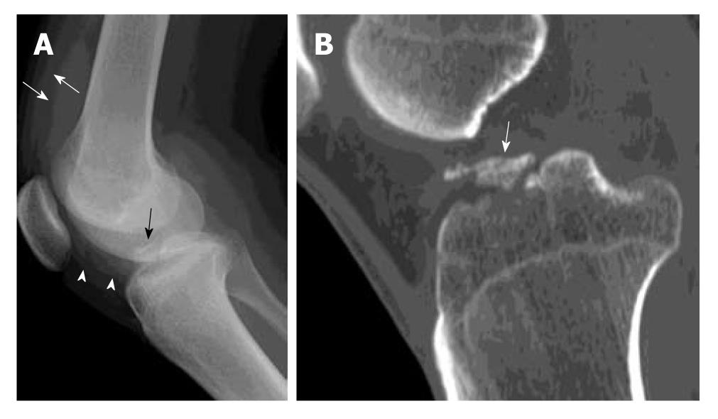
Figure 1 Avulsion fracture of tibial spine.
A 20-year-old man suffered knee injury during a football match. A: Lateral radiograph of the knee shows a displaced avulsion fracture of the anterior cruciate ligament (black arrow) at the anterior intercondylar eminence of tibia. The fracture fragment is completely elevated from the native bone. Increased soft tissue opacity in the suprapatellar pouch (white arrows) and infrapatellar pouch (white arrowheads) is in keeping with haemarthrosis; B: Reformatted sagittal computed tomography image of the same patient through the mid tibial plateau shows a displaced avulsion fracture of the tibial intercondylar eminence (white arrow).
- Citation: Ng WHA, Griffith JF, Hung EHY, Paunipagar B, Law BKY, Yung PSH. Imaging of the anterior cruciate ligament. World J Orthop 2011; 2(8): 75-84
- URL: https://www.wjgnet.com/2218-5836/full/v2/i8/75.htm
- DOI: https://dx.doi.org/10.5312/wjo.v2.i8.75


