Copyright
©2011 Baishideng Publishing Group Co.
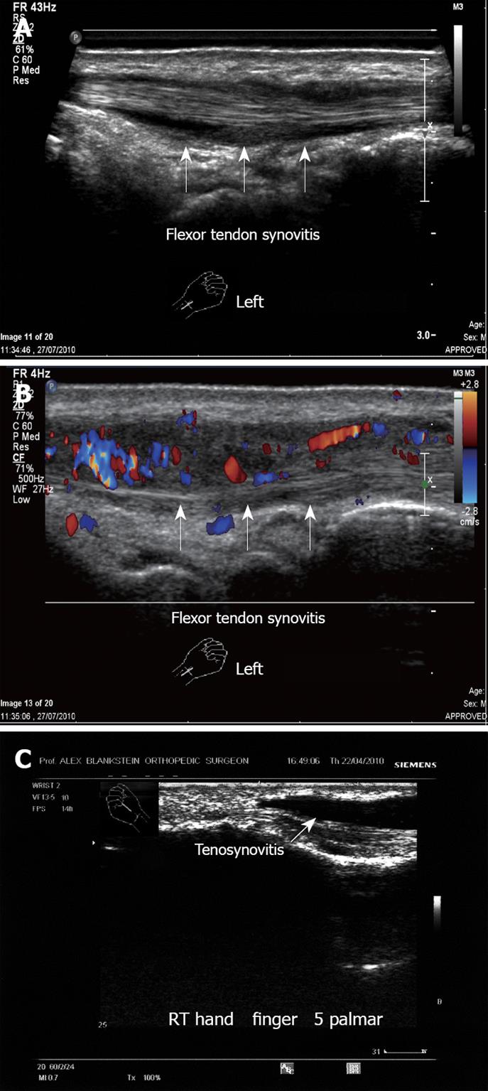
Figure 15 Tenosynovitis.
A: Left hand flexor tendon synovitis. Note the fluid around the tendon. No tear is demonstrated; B: Left hand flexor tendon synovitis. Note the hyper-vascularity with the vascular inflammation signs; C: Flexor tendon synovitis, right hand mid-phalanx. Note the large amount of clear fluid around the tendon. This 55-year old woman presented with acute pain in the palmar aspect of the hand with irregular synovial thickening, increased fluid and hypervascularity.
- Citation: Blankstein A. Ultrasound in the diagnosis of clinical orthopedics: The orthopedic stethoscope. World J Orthop 2011; 2(2): 13-24
- URL: https://www.wjgnet.com/2218-5836/full/v2/i2/13.htm
- DOI: https://dx.doi.org/10.5312/wjo.v2.i2.13


