Copyright
©The Author(s) 2025.
World J Orthop. Apr 18, 2025; 16(4): 102506
Published online Apr 18, 2025. doi: 10.5312/wjo.v16.i4.102506
Published online Apr 18, 2025. doi: 10.5312/wjo.v16.i4.102506
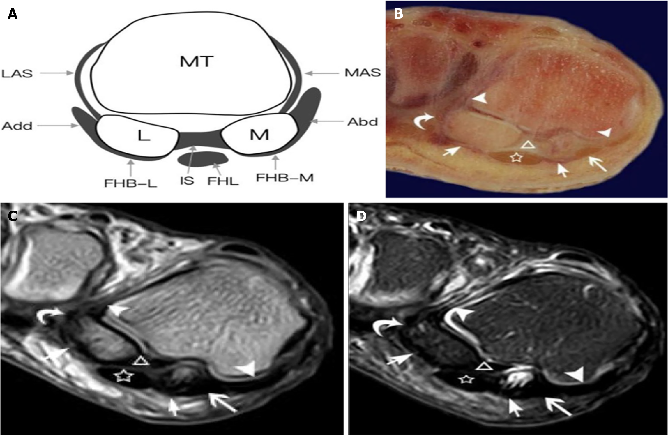
Figure 2 A 45-year-old right foot specimen revealing the capsuloligamentous complex of the first metatarsophalangeal joint[22].
Citation: Wang JE, Bai RJ, Zhan HL, Li WT, Qian ZH, Wang NL, Yin Y. High-resolution 3T magnetic resonance imaging and histological analysis of capsuloligamentous complex of the first metatarsophalangeal joint. J Orthop Surg Res 2021; 16: 638. Copyright© The Author(s) 2021. Published by Springer Nature. This image is licensed under a Creative Commons Attribution 4.0 International License, which permits use, sharing, adaptation, distribution, and reproduction in any medium, provided the original author(s) and source are credited. License details: https://creativecommons.org/Licenses/by/4.0/. A: Schematic coronal diagram through of first metatarsophalangeal joint and the sesamoid bones; B: Coronal anatomic section; C: and D: T1-weighted and T2-weighted spectral attenuated inversion recovery magnetic resonance imaging of the foot. LAS: Lateral accessory sesamoid ligament; Add: Adductor hallucis tendon; MT: Metatarsal; MAS: Medial accessory sesamoid ligamen; Adb: Abductor hallucis tendon; L: Lateral sesamoid; M: Medial sesamoid; FHB-L: Lateral head of flexor hallucis brevis tendon; IS: Intersesamoid ligament; FHL: Flexor hallucis longus tendon; FHB-M: Medial head of flexor hallucis brevis tendon; White triangle: The intersesamoid ligament; White pentagram: Tendon of flexor hallucis longus; White arrowheads: The accessory sesamoid ligaments; White short arrows: The flexor hallucis brevis tendons both medial and lateral heads; White long arrow; White curved arrow: The abductor hallucis and adductor hallucis tendons; White circle: The medial sesamoid phalangeal ligament.
- Citation: Embaby OM, Elalfy MM. First metatarsophalangeal joint: Embryology, anatomy and biomechanics. World J Orthop 2025; 16(4): 102506
- URL: https://www.wjgnet.com/2218-5836/full/v16/i4/102506.htm
- DOI: https://dx.doi.org/10.5312/wjo.v16.i4.102506


