Copyright
©The Author(s) 2025.
World J Orthop. Mar 18, 2025; 16(3): 101073
Published online Mar 18, 2025. doi: 10.5312/wjo.v16.i3.101073
Published online Mar 18, 2025. doi: 10.5312/wjo.v16.i3.101073
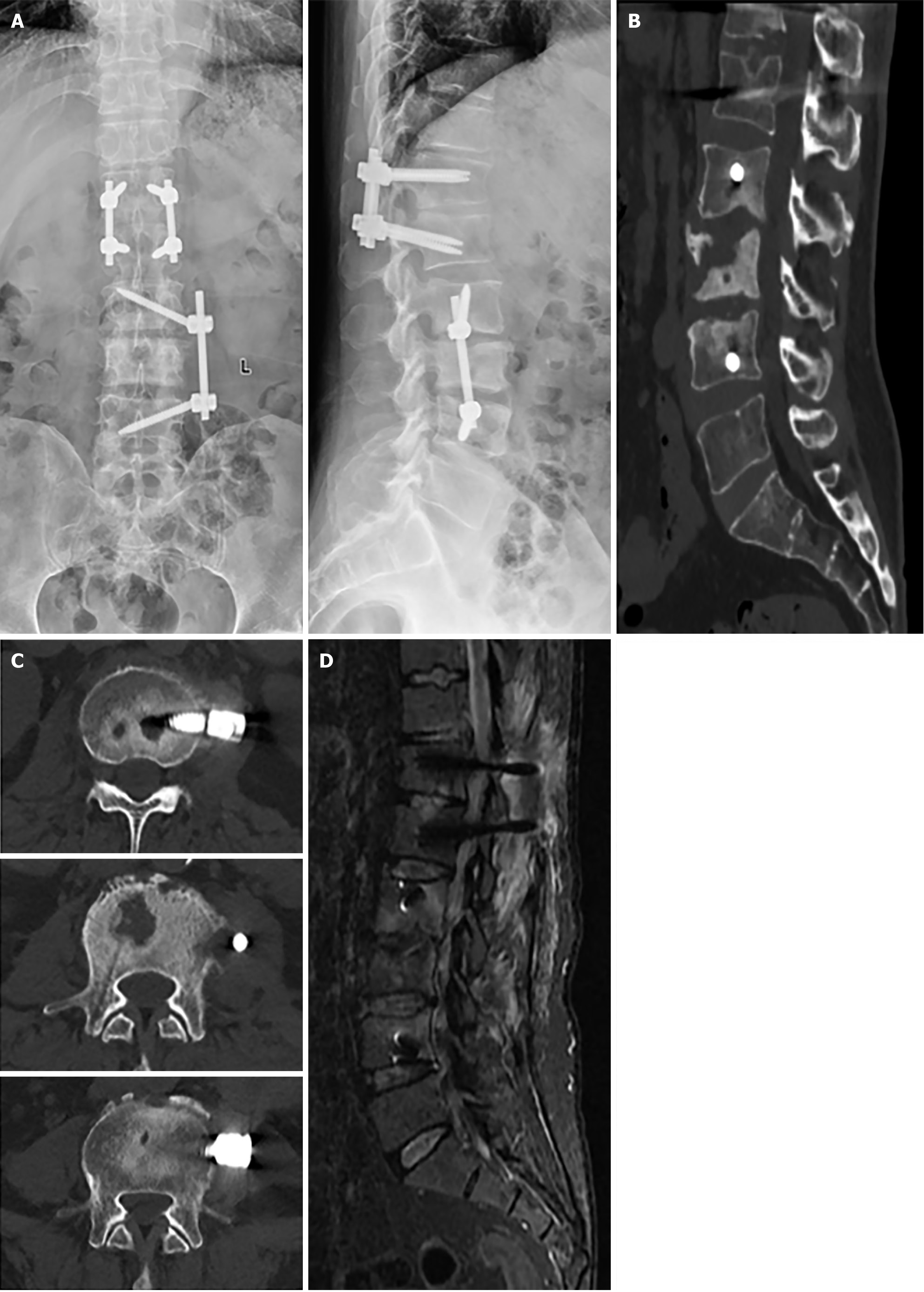
Figure 5 Lumbar spine imaging at 3 months postoperatively.
A: Anteroposterior and lateral radiography of the lumbar spine; B: Sagittal computed tomography (CT) of the lumbar spine; C: Axial CT of the lumbar spine, demonstrating destructed vertebral bodies in L2–L4; D: Sagittal T2-weighted fat suppression magnetic resonance imaging showing resolution of abnormal signals in the vertebral bodies and intervertebral spaces disappeared compared with preoperative imaging.
- Citation: Wu JJ, Chang ZQ. Treatment of refractory thoracolumbar spine infection by thirteen times of vacuum sealing drainage: A case report. World J Orthop 2025; 16(3): 101073
- URL: https://www.wjgnet.com/2218-5836/full/v16/i3/101073.htm
- DOI: https://dx.doi.org/10.5312/wjo.v16.i3.101073


