Copyright
©The Author(s) 2025.
World J Orthop. Feb 18, 2025; 16(2): 98674
Published online Feb 18, 2025. doi: 10.5312/wjo.v16.i2.98674
Published online Feb 18, 2025. doi: 10.5312/wjo.v16.i2.98674
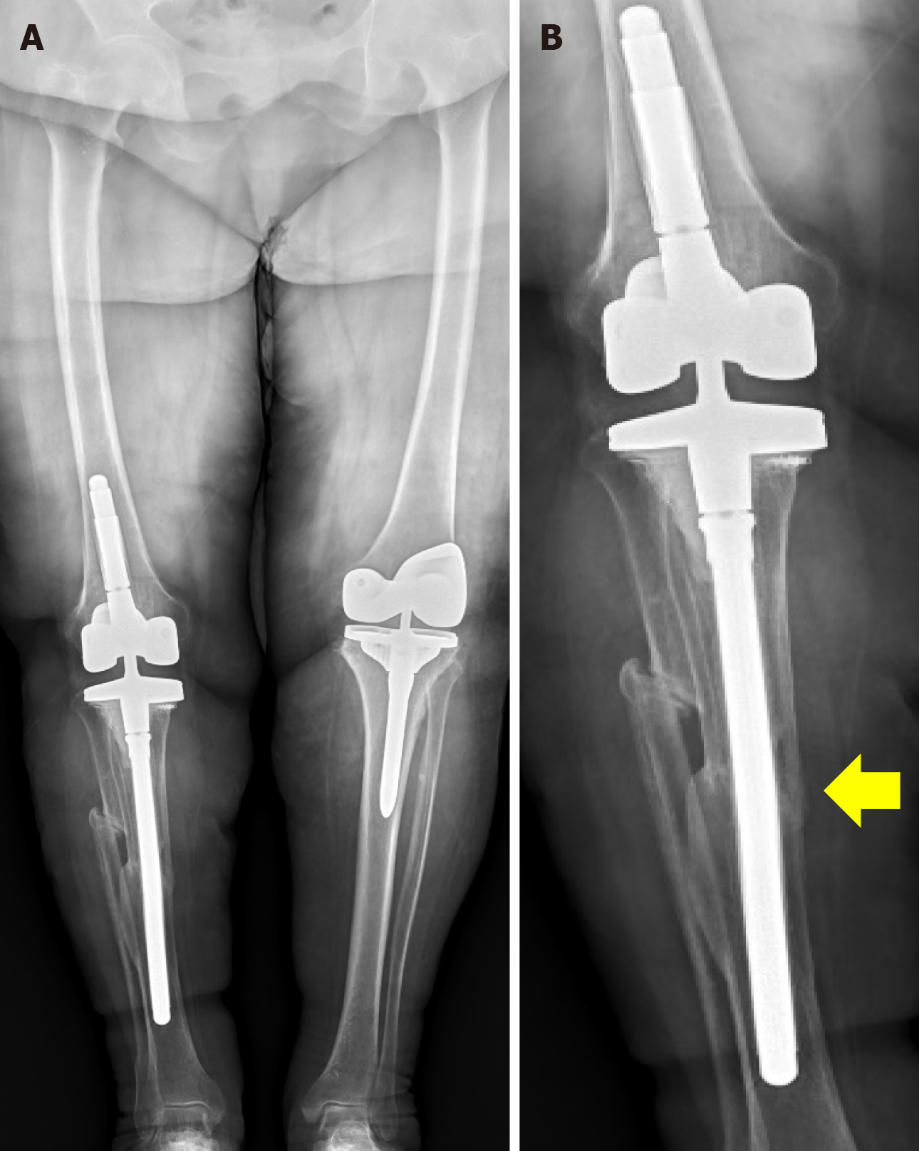
Figure 5 Radiographs 2 years after revision total knee arthroplasty of the right knee, and 6 months after left total knee arthroplasty.
A: Standing long leg; B: Plain anteroposterior. Full bone union of the right tibia was confirmed (yellow arrow). Distally, progressive loss of the anterior tibial cortex was revealed as a result of the load exerted by the distal part of stem.
- Citation: Kocon M, Grzelecki D. Periprosthetic fractures of the tibial shaft following long-stemmed total knee arthroplasty: A case report. World J Orthop 2025; 16(2): 98674
- URL: https://www.wjgnet.com/2218-5836/full/v16/i2/98674.htm
- DOI: https://dx.doi.org/10.5312/wjo.v16.i2.98674


