Copyright
©The Author(s) 2025.
World J Orthop. Feb 18, 2025; 16(2): 104095
Published online Feb 18, 2025. doi: 10.5312/wjo.v16.i2.104095
Published online Feb 18, 2025. doi: 10.5312/wjo.v16.i2.104095
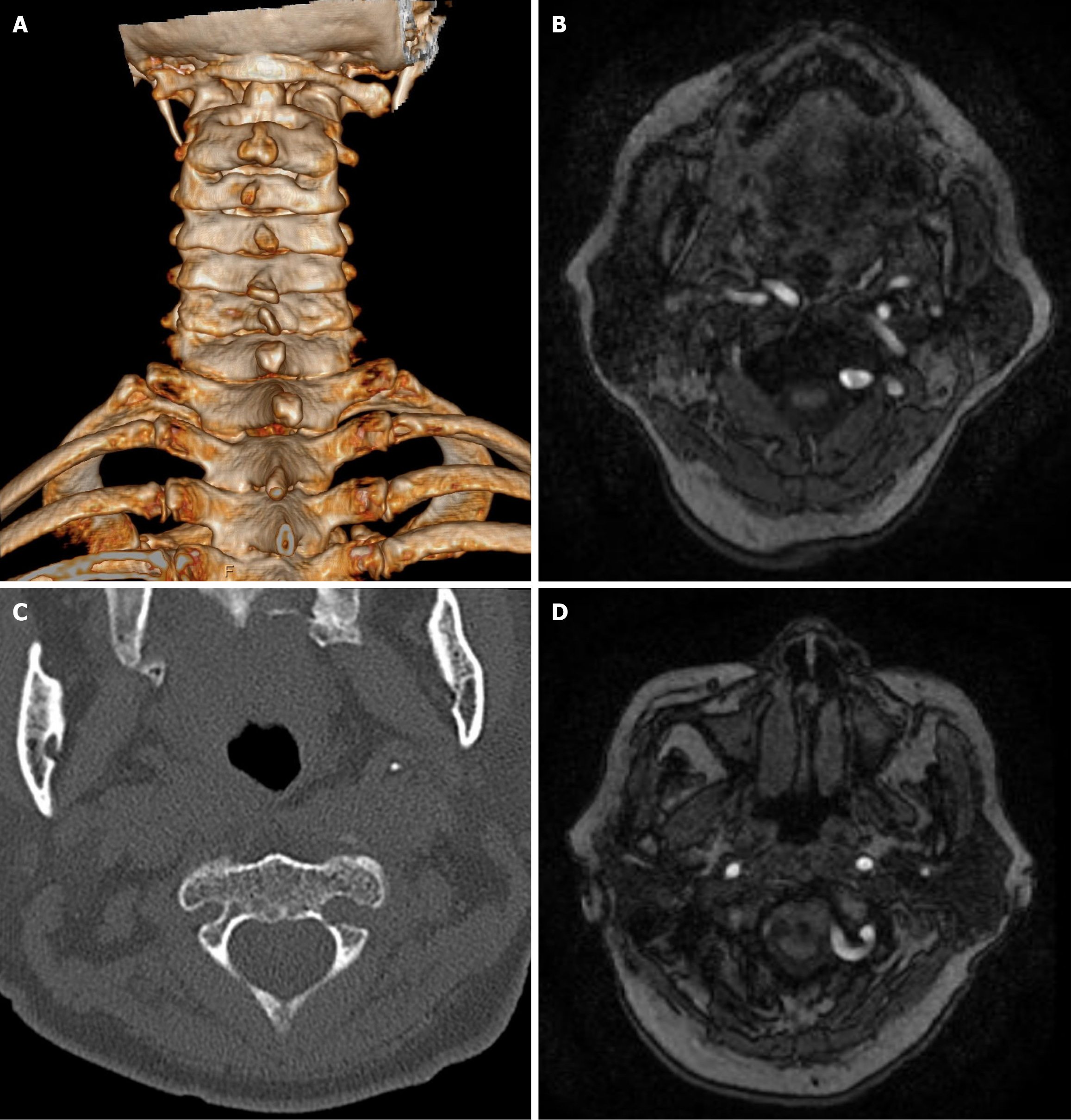
Figure 3 Preoperative computed tomography reconstruction and magnetic resonance tomography images of the patient.
A: Computed tomography (CT) reconstruction scans demonstrated spontaneous atlanto-occipital fusion; B-D: CT cross-sectional images indicate a high-riding vertebral artery anomaly and restricted placement of screws on the left side of the C2 vertebra. Vascular MRI imaging indicates invasion of the left vertebral artery into the lateral mass of C2 and the left posterior arch of C1.
- Citation: Tang RH, Yin J, Zhou ZW. Atlantoaxial dislocation with vertebral artery anomaly: A case report. World J Orthop 2025; 16(2): 104095
- URL: https://www.wjgnet.com/2218-5836/full/v16/i2/104095.htm
- DOI: https://dx.doi.org/10.5312/wjo.v16.i2.104095


