Copyright
©The Author(s) 2025.
World J Orthop. Feb 18, 2025; 16(2): 103416
Published online Feb 18, 2025. doi: 10.5312/wjo.v16.i2.103416
Published online Feb 18, 2025. doi: 10.5312/wjo.v16.i2.103416
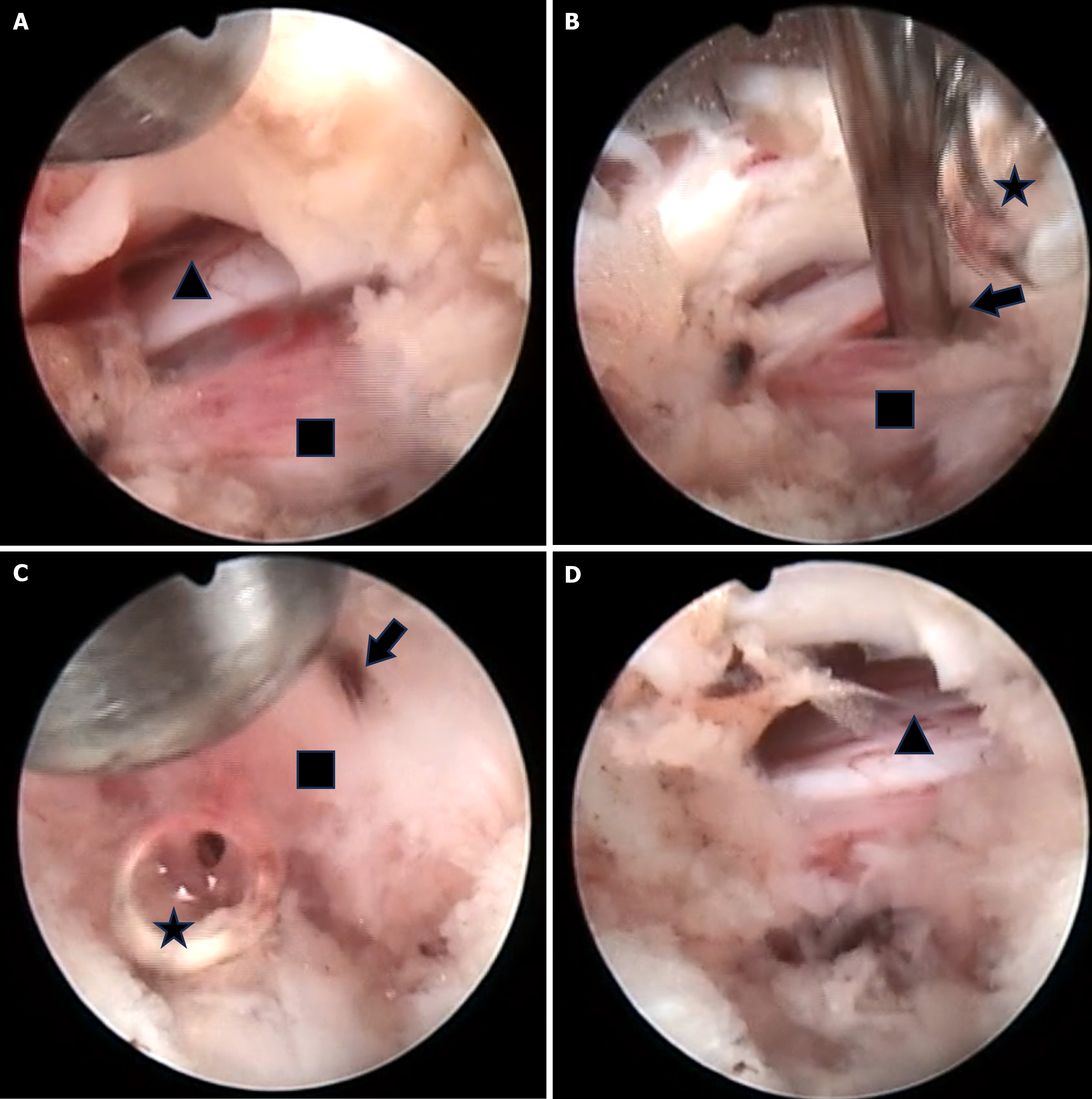
Figure 4 Intraoperative endoscopic images.
A: The L5 nerve root was compressed by a pseudocyst; B: Air bubbles observed when the pseudocyst was punctured; C: Removing the pseudocyst wall using straight and flexible graspers; D: The pseudocyst was completely removed and the nerve root was decompressed. Square: Gas-containing pseudocyst; Triangle: L5 nerve root; Star: Air bubbles; Arrow: Fluid flowing out from the pseudocyst.
- Citation: Jiang ZX, Ren JB, Li YC, Chen L. Percutaneous transforaminal endoscopic treatment of gas-containing pseudocyst compressing the L5 nerve root: A case report. World J Orthop 2025; 16(2): 103416
- URL: https://www.wjgnet.com/2218-5836/full/v16/i2/103416.htm
- DOI: https://dx.doi.org/10.5312/wjo.v16.i2.103416


