Copyright
©The Author(s) 2025.
World J Orthop. Jan 18, 2025; 16(1): 101424
Published online Jan 18, 2025. doi: 10.5312/wjo.v16.i1.101424
Published online Jan 18, 2025. doi: 10.5312/wjo.v16.i1.101424
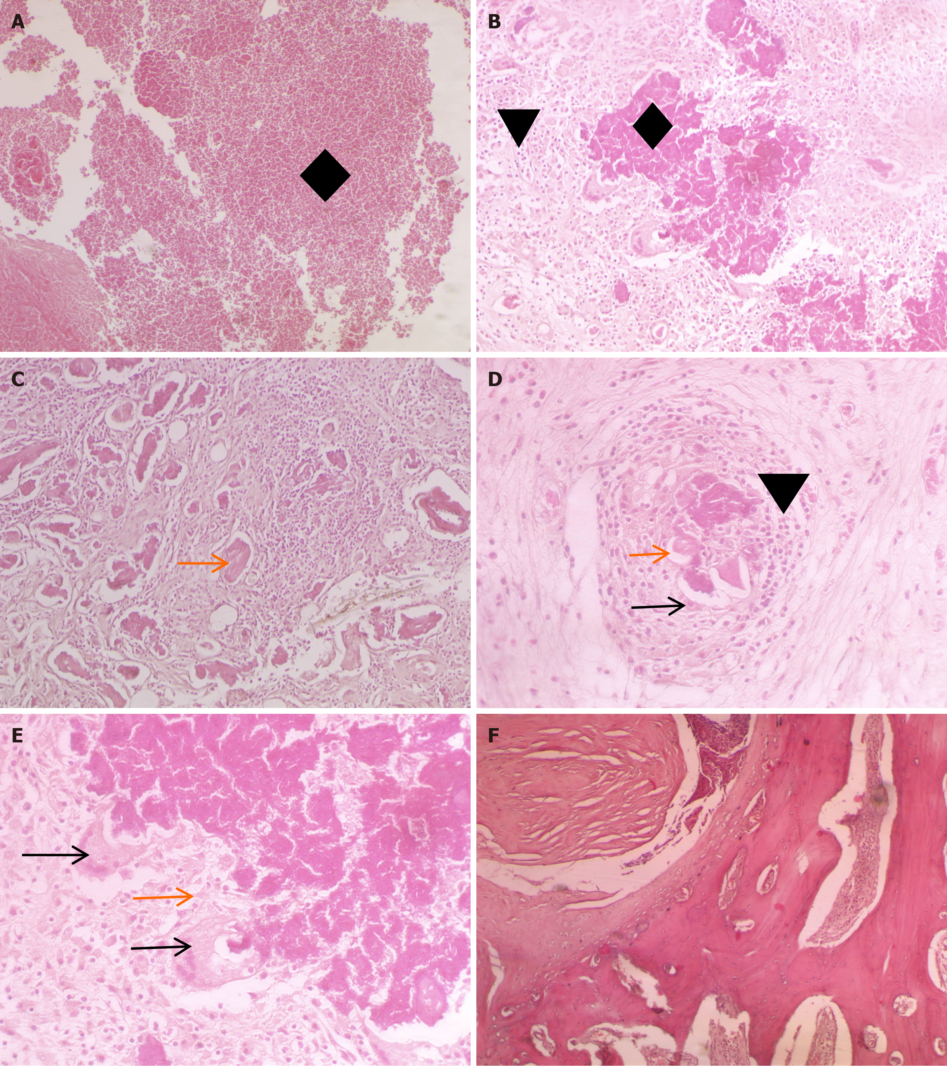
Figure 3 Twelve-week post-operative histopathological results using hematoxylin and eosin stain.
A: Pathological changes typical of tuberculosis with vast areas of caseous necrosis [diamond; hematoxylin and eosin (HE) × 200]; B: Infiltration of inflammatory cells (triangle) and caseous necrosis (diamond; HE × 200); C: Disordered trabecular bone structure and numerous sequestra (orange arrow; HE × 100); D: Typical tuberculous nodules with caseous necrotic material in the center: Epithelioid cells (orange arrow), multinucleated giant cells (black arrow) and inflammatory cell infiltration (triangle; HE × 400); E: Typical multinucleated giant cells (black arrow) and epithelioid cells (orange arrow) were observed (HE × 400); F: No abnormalities were observed in the control group (HE × 200).
- Citation: Qiao YJ, Song XY, Zhang LD, Li F, Zhang HQ, Zhou SH. Comparative study of a rabbit model of spinal tuberculosis using different concentrations of Mycobacterium tuberculosis. World J Orthop 2025; 16(1): 101424
- URL: https://www.wjgnet.com/2218-5836/full/v16/i1/101424.htm
- DOI: https://dx.doi.org/10.5312/wjo.v16.i1.101424


