Copyright
©The Author(s) 2025.
World J Orthop. Jan 18, 2025; 16(1): 101424
Published online Jan 18, 2025. doi: 10.5312/wjo.v16.i1.101424
Published online Jan 18, 2025. doi: 10.5312/wjo.v16.i1.101424
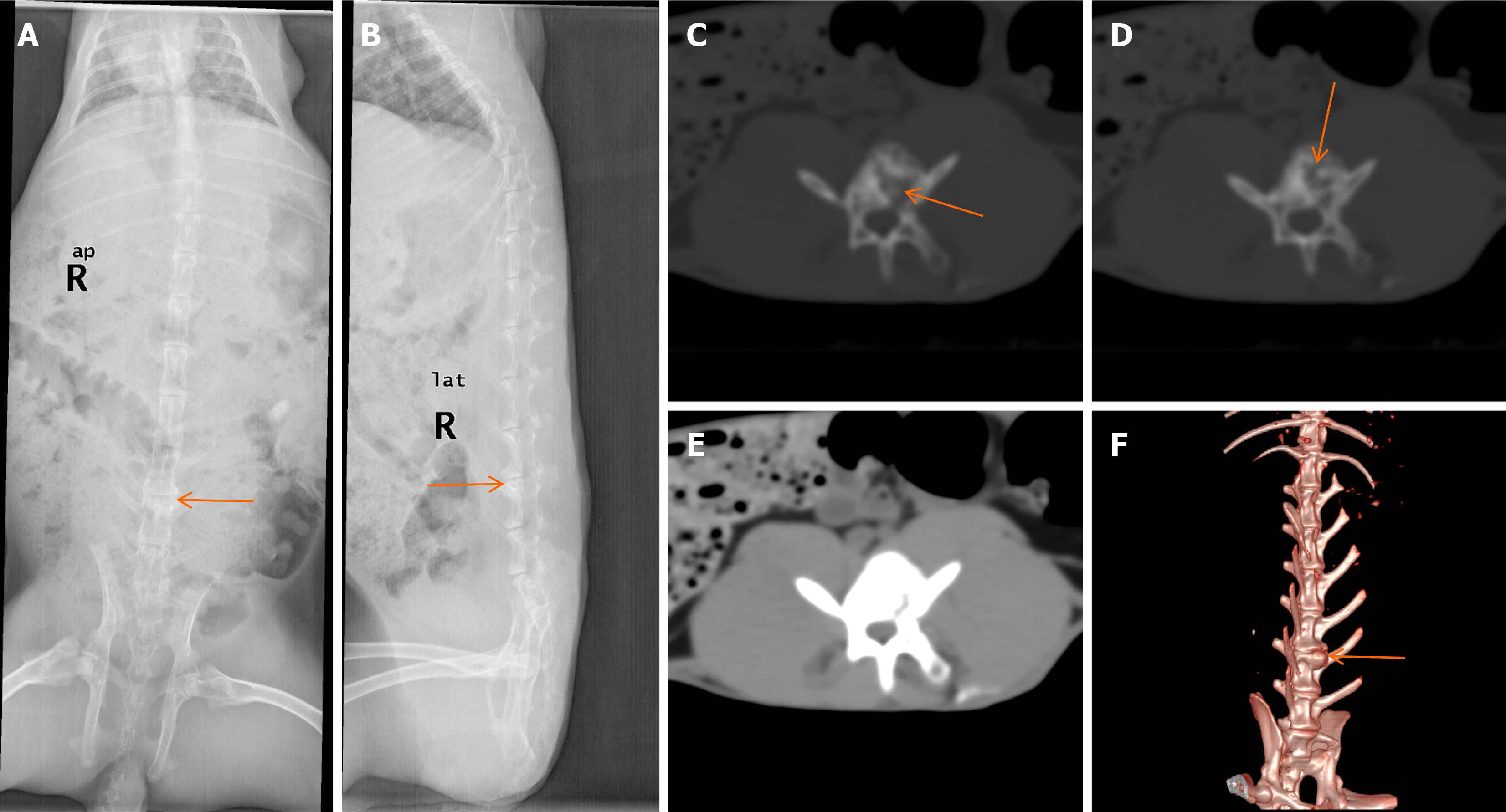
Figure 2 Twelve-week post-operative imaging results.
A and B: X-ray analysis revealed that the intervertebral spaces appeared blurred and narrowed, the adjacent endplates appeared to have increased bone density shadows (orange arrows); C-E: Computed tomography (CT) scans showed that dotted sequestra and increased bone mineral densities shadows could be observed in the vertebral bodies; the local vertebral bone cortices were nonunion (orange arrows). There was no obvious soft tissue swelling around the vertebral bodies; F: 3-dimensional CT scans showed that the intervertebral spaces had become narrowed with surrounding osteophyte formation (orange arrows).
- Citation: Qiao YJ, Song XY, Zhang LD, Li F, Zhang HQ, Zhou SH. Comparative study of a rabbit model of spinal tuberculosis using different concentrations of Mycobacterium tuberculosis. World J Orthop 2025; 16(1): 101424
- URL: https://www.wjgnet.com/2218-5836/full/v16/i1/101424.htm
- DOI: https://dx.doi.org/10.5312/wjo.v16.i1.101424


