Copyright
©The Author(s) 2024.
World J Orthop. Sep 18, 2024; 15(9): 891-901
Published online Sep 18, 2024. doi: 10.5312/wjo.v15.i9.891
Published online Sep 18, 2024. doi: 10.5312/wjo.v15.i9.891
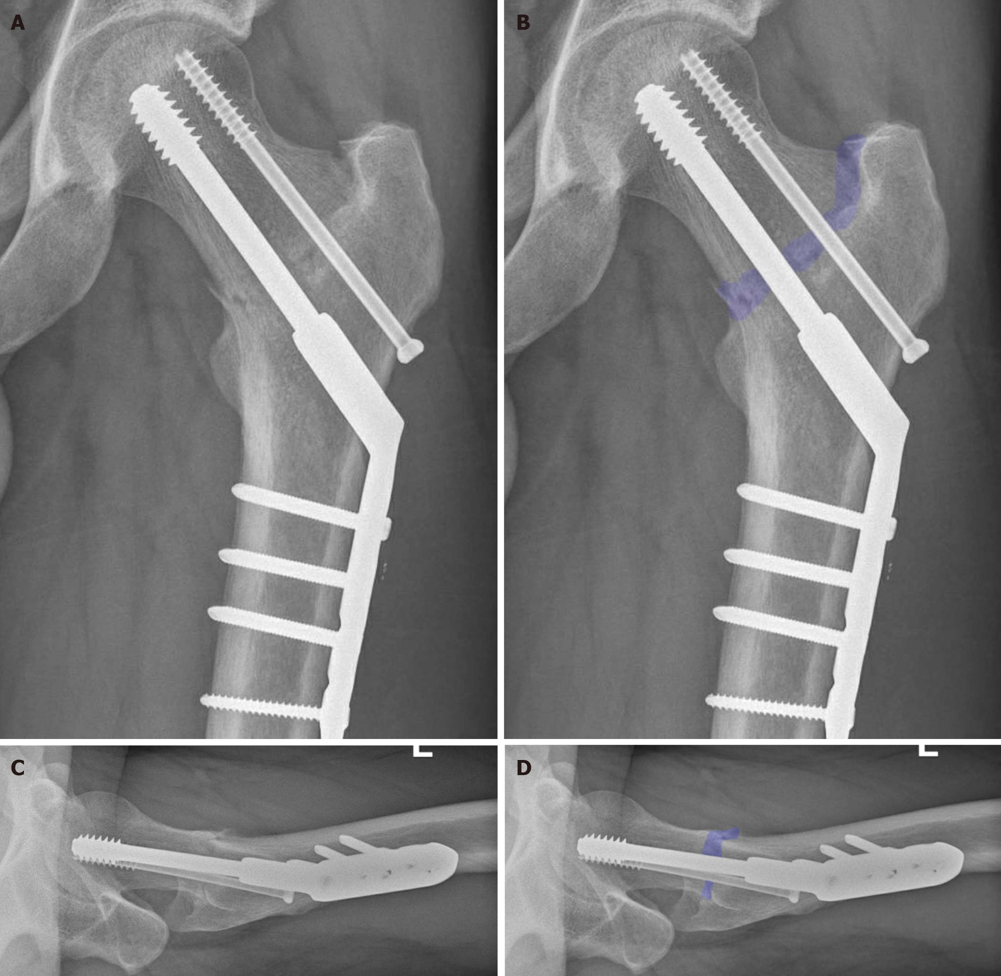
Figure 7 Position of osteosynthesis material at outpatient clinic follow-up.
A: Coronal X-ray without overlay; B: Coronal X-ray including an overlay, highlighting the fracture; C: Lateral X-ray without overlay; D: Lateral X-ray including an overlay, highlighting the fracture. During the outpatient clinic follow-up, the patient was almost free of complaints and the alignment was consistent with before. At this time, there were no convincing signs of bone remodeling, which is also not expected with this atypical fracture.
- Citation: Oudmaijer CA, Paulino Pereira NR, Visser D, Wakker AM, Veltman ES, van Linschoten R. Lateral femoral neck stress fractures: A case report. World J Orthop 2024; 15(9): 891-901
- URL: https://www.wjgnet.com/2218-5836/full/v15/i9/891.htm
- DOI: https://dx.doi.org/10.5312/wjo.v15.i9.891


