Copyright
©The Author(s) 2024.
World J Orthop. Sep 18, 2024; 15(9): 891-901
Published online Sep 18, 2024. doi: 10.5312/wjo.v15.i9.891
Published online Sep 18, 2024. doi: 10.5312/wjo.v15.i9.891
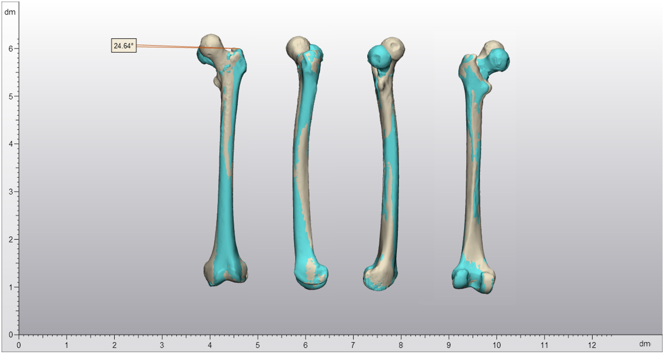
Figure 4 3D reconstruction of the fracture and malalignment at presentation.
This 3D reconstruction illustrates the malalignment: 24.6° of external rotation and 16.0 mm shortening due to varus alignment at the fracture site. From left to right: Images from anterior, lateral, medial, and posterior perspectives. The sand-colored reconstruction represents the contralateral side; cyan, the affected side.
- Citation: Oudmaijer CA, Paulino Pereira NR, Visser D, Wakker AM, Veltman ES, van Linschoten R. Lateral femoral neck stress fractures: A case report. World J Orthop 2024; 15(9): 891-901
- URL: https://www.wjgnet.com/2218-5836/full/v15/i9/891.htm
- DOI: https://dx.doi.org/10.5312/wjo.v15.i9.891


