Copyright
©The Author(s) 2024.
World J Orthop. Aug 18, 2024; 15(8): 683-695
Published online Aug 18, 2024. doi: 10.5312/wjo.v15.i8.683
Published online Aug 18, 2024. doi: 10.5312/wjo.v15.i8.683
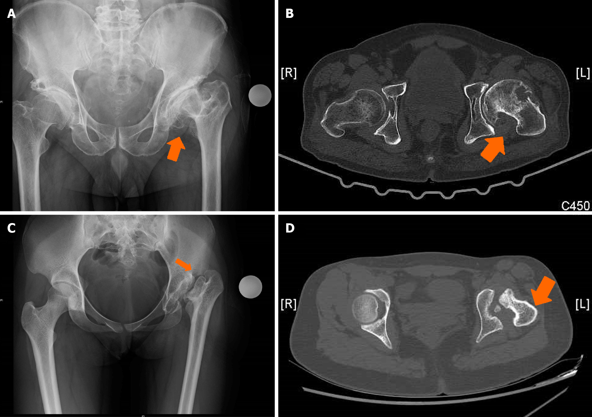
Figure 4 Anatomical abnormalities in the proximal femur include anteversion changes.
A and B: X-ray of both hips anteroposterior (AP) showing dysplastic hip left with computed tomography (CT) assessment showing increased anteversion and deformed head and acetabulum; C and D: X-ray of both hips AP showing postseptic sequelae in left hip at total hip arthroplasty with computed tomography (CT) images showing large medial osteophyte, head malformation and increased femoral anteversion. Arrows indicating the changes in the X-ray and CT images. Preoperative evaluation helps in identification and adjustment of intraoperative femoral version.
- Citation: Oommen AT. Total hip arthroplasty for sequelae of childhood hip disorders: Current review of management to achieve hip centre restoration. World J Orthop 2024; 15(8): 683-695
- URL: https://www.wjgnet.com/2218-5836/full/v15/i8/683.htm
- DOI: https://dx.doi.org/10.5312/wjo.v15.i8.683


