Copyright
©The Author(s) 2024.
World J Orthop. Jul 18, 2024; 15(7): 642-649
Published online Jul 18, 2024. doi: 10.5312/wjo.v15.i7.642
Published online Jul 18, 2024. doi: 10.5312/wjo.v15.i7.642
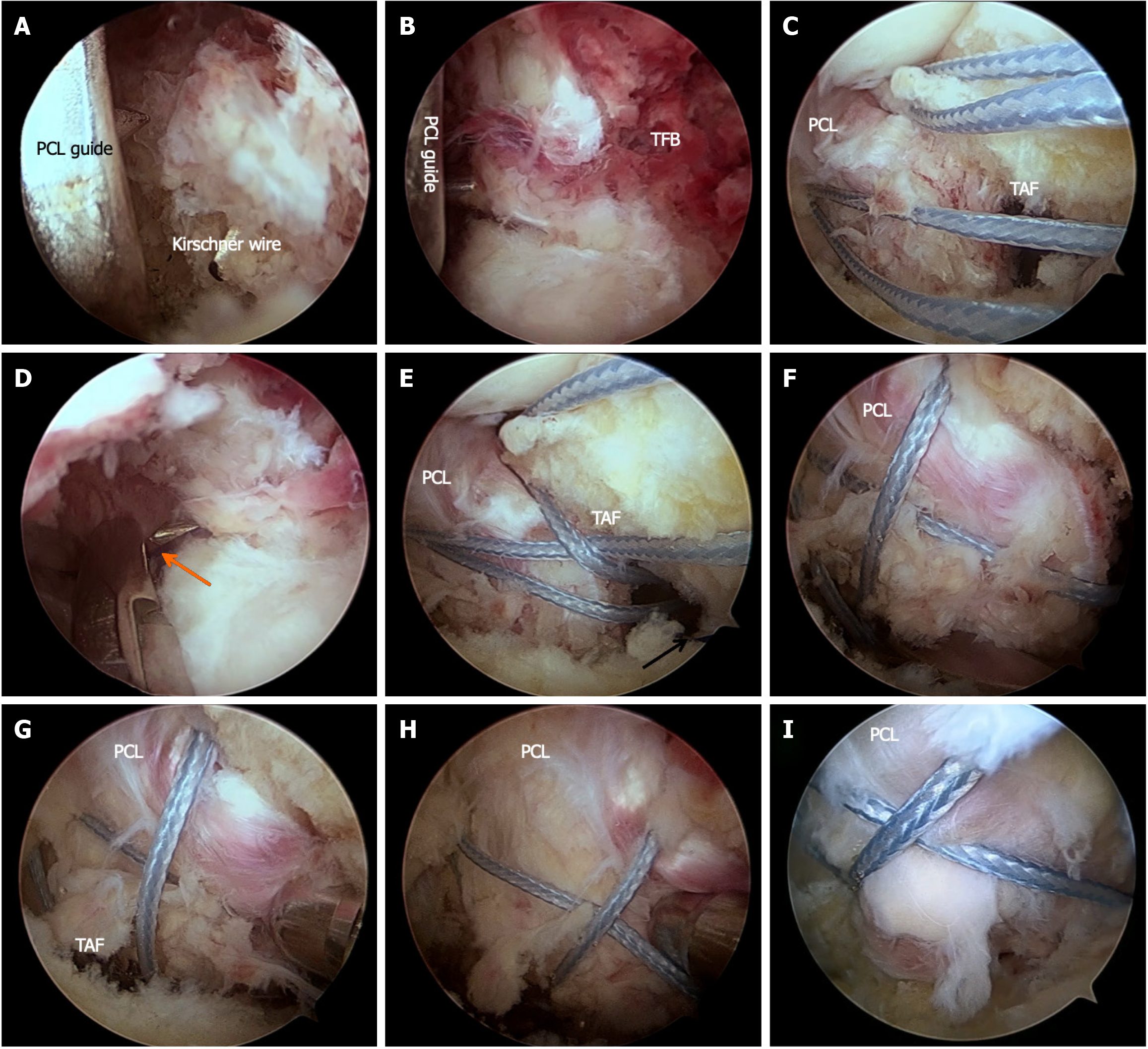
Figure 1 Intraoperative arthroscopic views.
A and B: The tips of the guide were placed at the inferolateral or inferomedial corners of the tibial fracture bed. A 2.5 mm Kirschner guide wire was drilled to established two tibial tunnels; C: Two Ethicon MB66 sutures were introduced by polydioxanone (PDS) suture; D: An epidural needle was used to introduce a PDS suture through one of the tibial tunnels; E: Use the PDS suture to draw out both ends of one of the Ethicon MB66 sutures to the tibial tunnel opening; F: A suture grasper was used to adjust the position of the two Ethicon MB66 sutures so that the two sutures pressed on the fracture fragment in an "M" shape; G and H: The bone fragments were reduced with the assistance of a probe; I: The reduction of avulsion fracture was achieved with the tightened suture. PCL: Posterior cruciate ligament; TFB: Tibial fracture bed; TAF: Tibia avulsion fracture; PDS: Polydioxanone.
- Citation: Zhang XH, Yu J, Zhao MY, Cao JH, Wu B, Xu DF. Arthroscopic M-shaped suture fixation for tibia avulsion fracture of posterior cruciate ligament: A modified technique and case series. World J Orthop 2024; 15(7): 642-649
- URL: https://www.wjgnet.com/2218-5836/full/v15/i7/642.htm
- DOI: https://dx.doi.org/10.5312/wjo.v15.i7.642


