Copyright
©The Author(s) 2024.
World J Orthop. Jun 18, 2024; 15(6): 593-601
Published online Jun 18, 2024. doi: 10.5312/wjo.v15.i6.593
Published online Jun 18, 2024. doi: 10.5312/wjo.v15.i6.593
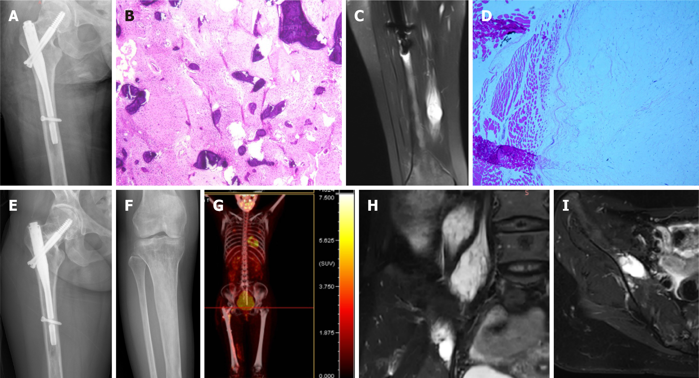
Figure 3 Representative images.
A and B: X-ray image (A) of the right femur after internal fixation, and representative hematoxylin-eosin (HE) staining (B) of postoperative specimen from the femoral fibrous dysplasia; C and D: Representative magnetic resonance (MR) image (C) and HE staining (D) of the intramuscular myxoma in right vastus medialis; E-I: Results of 1-year follow-up of femur X-ray, iliopsoas myxoma and iliac crest fibrous dysplasia. Representative images of femur (E) and tibia (F) X-ray, positron emission tomography-CT (G), iliopsoas myxoma MR (H), and iliac fibrous dysplasia MR (I) at 1-year follow-up.
- Citation: Li XM, Chen ZH, Wang KY, Chen JN, Yao ZN, Yao YH, Zhou XW, Lin N. Mazabraud’s syndrome in female patients: Two case reports. World J Orthop 2024; 15(6): 593-601
- URL: https://www.wjgnet.com/2218-5836/full/v15/i6/593.htm
- DOI: https://dx.doi.org/10.5312/wjo.v15.i6.593


