Copyright
©The Author(s) 2024.
World J Orthop. Jun 18, 2024; 15(6): 593-601
Published online Jun 18, 2024. doi: 10.5312/wjo.v15.i6.593
Published online Jun 18, 2024. doi: 10.5312/wjo.v15.i6.593
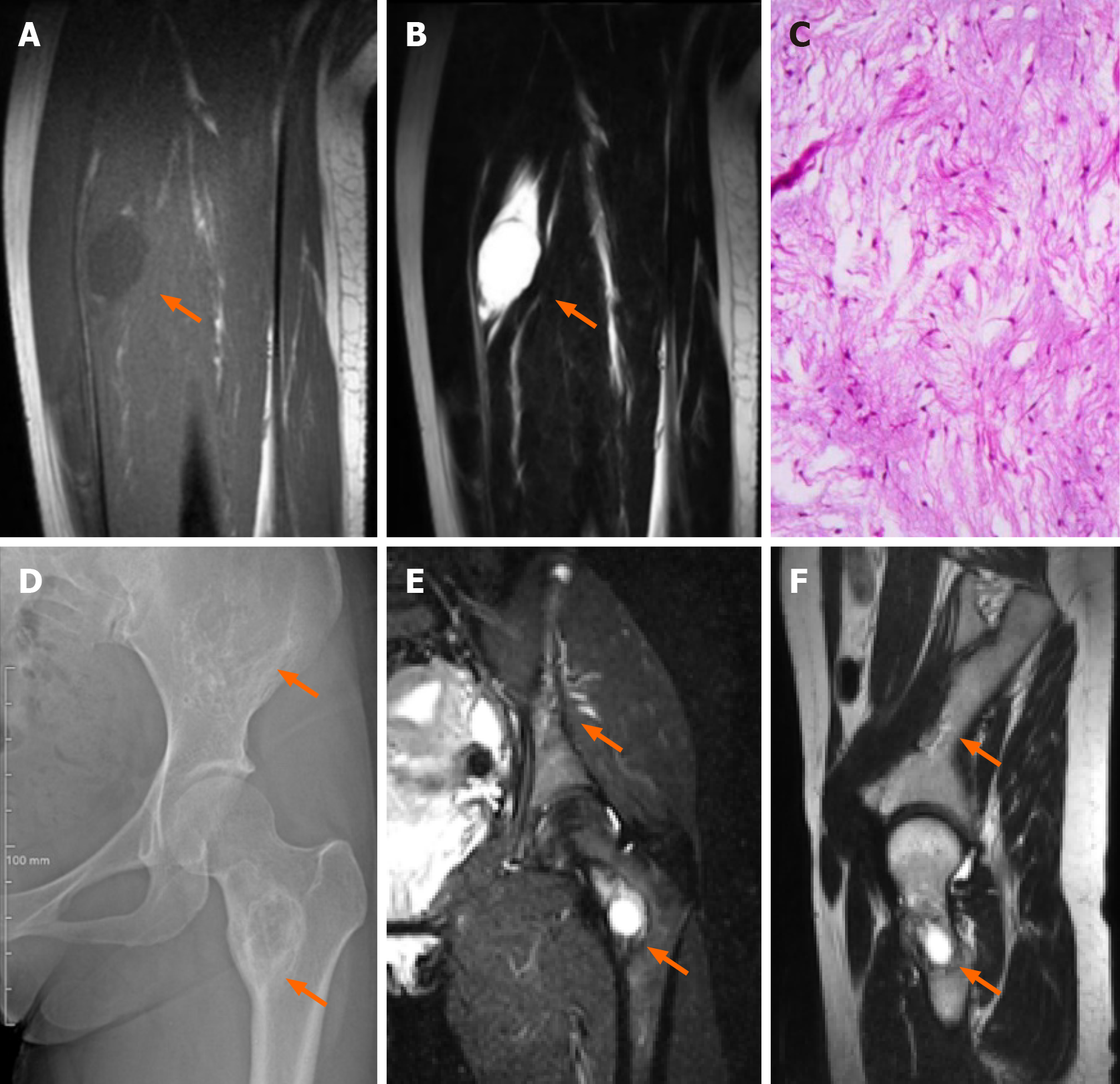
Figure 2 Representative images.
A-C: Representative T1WI (A) and T2WI (B) of MR images, and hematoxylin-eosin staining (C) of the intramuscular lesion (orange arrows) in vastus intermedius muscle; D-F: Representative X-ray (D), T2WI (E) and T1WI images (F) showing bone lesions (orange arrows) in the pelvis and femur.
- Citation: Li XM, Chen ZH, Wang KY, Chen JN, Yao ZN, Yao YH, Zhou XW, Lin N. Mazabraud’s syndrome in female patients: Two case reports. World J Orthop 2024; 15(6): 593-601
- URL: https://www.wjgnet.com/2218-5836/full/v15/i6/593.htm
- DOI: https://dx.doi.org/10.5312/wjo.v15.i6.593


