Copyright
©The Author(s) 2024.
World J Orthop. May 18, 2024; 15(5): 457-468
Published online May 18, 2024. doi: 10.5312/wjo.v15.i5.457
Published online May 18, 2024. doi: 10.5312/wjo.v15.i5.457
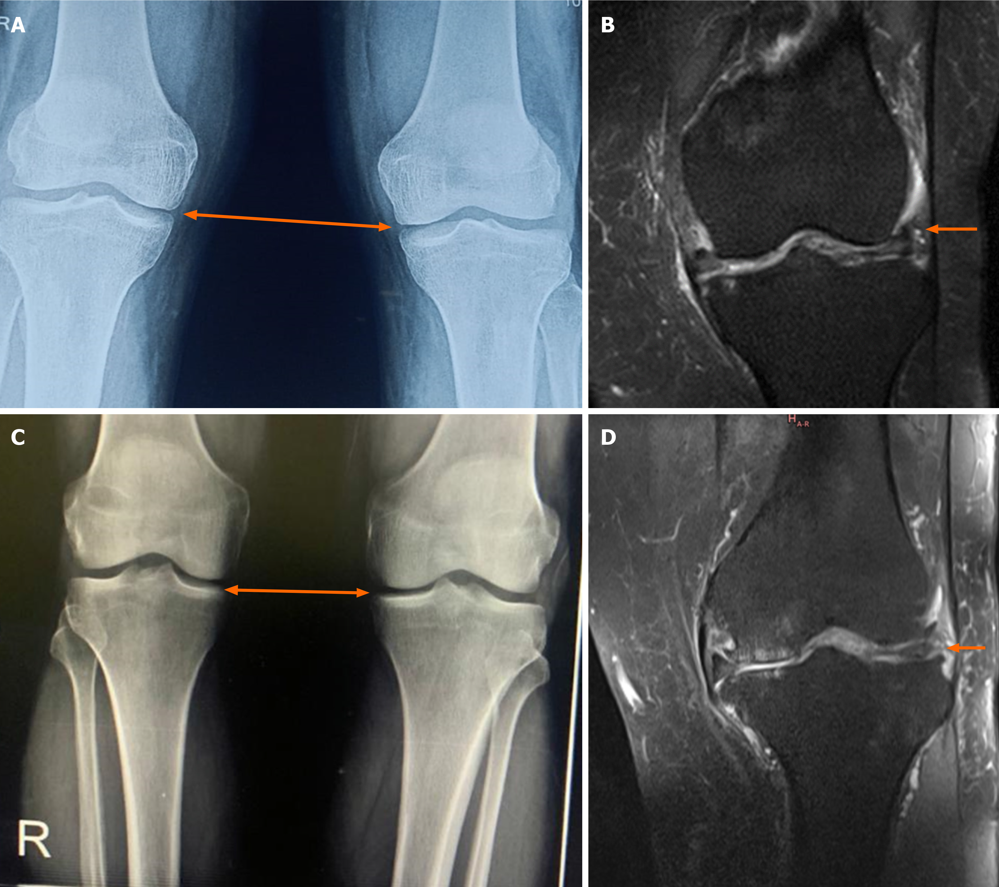
Figure 4 A representational case from group B.
A: Pre-procedural radiograph of bilateral knees (anteroposterior (AP) view on standing position) showing decreased medial joint line in bilateral knees suggestive of Kellgren Lawrence grade II knee osteoarthritis (OA); B: Pre-procedural T2W magnetic resonance imaging (MRI) (coronal section) showing hyperintensity with thinned out cartilage along the medial femoral condyle suggestive of OA knee; C: 2-years follow-up radiograph of bilateral knees (AP view on standing position) showing no improvement in the thickness of medial joint line in bilateral knees; D: 2-years follow-up T2W MRI (coronal image) showing no cartilaginous thickness indicating response to corticosteroids therapy.
- Citation: Jeyaraman M, Jeyaraman N, Jayakumar T, Ramasubramanian S, Ranjan R, Jha SK, Gupta A. Efficacy of stromal vascular fraction for knee osteoarthritis: A prospective, single-centre, non-randomized study with 2 years follow-up. World J Orthop 2024; 15(5): 457-468
- URL: https://www.wjgnet.com/2218-5836/full/v15/i5/457.htm
- DOI: https://dx.doi.org/10.5312/wjo.v15.i5.457


