Copyright
©The Author(s) 2024.
World J Orthop. Mar 18, 2024; 15(3): 238-246
Published online Mar 18, 2024. doi: 10.5312/wjo.v15.i3.238
Published online Mar 18, 2024. doi: 10.5312/wjo.v15.i3.238
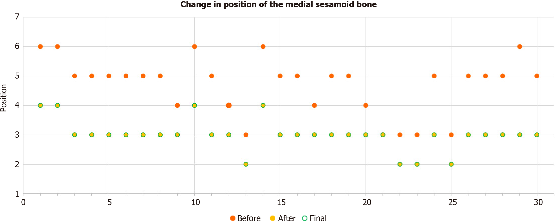
Figure 5 Change in position of the medial sesamoid bone for each operated foot.
Anteroposterior X-rays were graded using the Hardy and Clapham scale: (1) Position 1: The entire sesamoid is medial to the first metatarsal bisector; (2) Position 2: The lateral aspect of the sesamoid is tangential to the metatarsal bisector; (3) Position 3: The lateral one-third of the sesamoid overlaps the bisector; (4) Position 4: The sesamoid is centred over the bisector; (5) Position 5: The medial one-third of the sesamoid overlaps the bisector; (6) Position 6: The medial aspect of the sesamoid is tangential to the bisector; and (7) Position 7: The entire sesamoid is lateral to the bisector.
- Citation: Zhanaspayev A, Bokembayev N, Zhanaspayev M, Tlemissov A, Aubakirova S, Prokazyuk A. Correction method for moderate and severe degrees of hallux valgus associated with transfer metatarsalgia. World J Orthop 2024; 15(3): 238-246
- URL: https://www.wjgnet.com/2218-5836/full/v15/i3/238.htm
- DOI: https://dx.doi.org/10.5312/wjo.v15.i3.238


