Copyright
©The Author(s) 2024.
World J Orthop. Dec 18, 2024; 15(12): 1175-1182
Published online Dec 18, 2024. doi: 10.5312/wjo.v15.i12.1175
Published online Dec 18, 2024. doi: 10.5312/wjo.v15.i12.1175
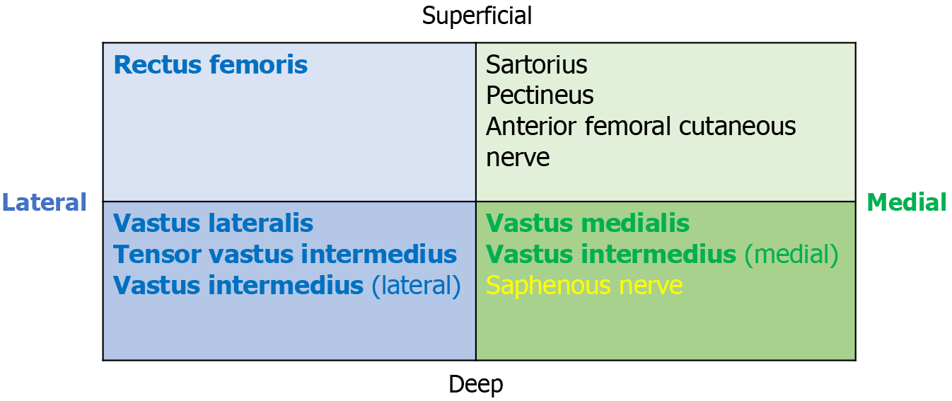
Figure 4 Schematic cross-section of the femoral nerve.
It consists of medial and lateral areas subdivided into superficial and deep portions. The rectus femoris, vastus lateralis, tensor vastus intermedius, and lateral area of the vastus intermedius are located in the lateral division (blue). The medial area of the vastus intermedius and the vastus medialis are located in the medial division (green). The anterior femoral cutaneous nerve and saphenous nerve are sensory nerve branches, and the sartorius and pectineus muscles are supplied by the superficial medial division.
- Citation: Spuehler D, Kuster L, Ullrich O, Grob K. Femoral nerve palsy following Girdlestone resection arthroplasty: An observational cadaveric study. World J Orthop 2024; 15(12): 1175-1182
- URL: https://www.wjgnet.com/2218-5836/full/v15/i12/1175.htm
- DOI: https://dx.doi.org/10.5312/wjo.v15.i12.1175


