Copyright
©The Author(s) 2024.
World J Orthop. Dec 18, 2024; 15(12): 1118-1123
Published online Dec 18, 2024. doi: 10.5312/wjo.v15.i12.1118
Published online Dec 18, 2024. doi: 10.5312/wjo.v15.i12.1118
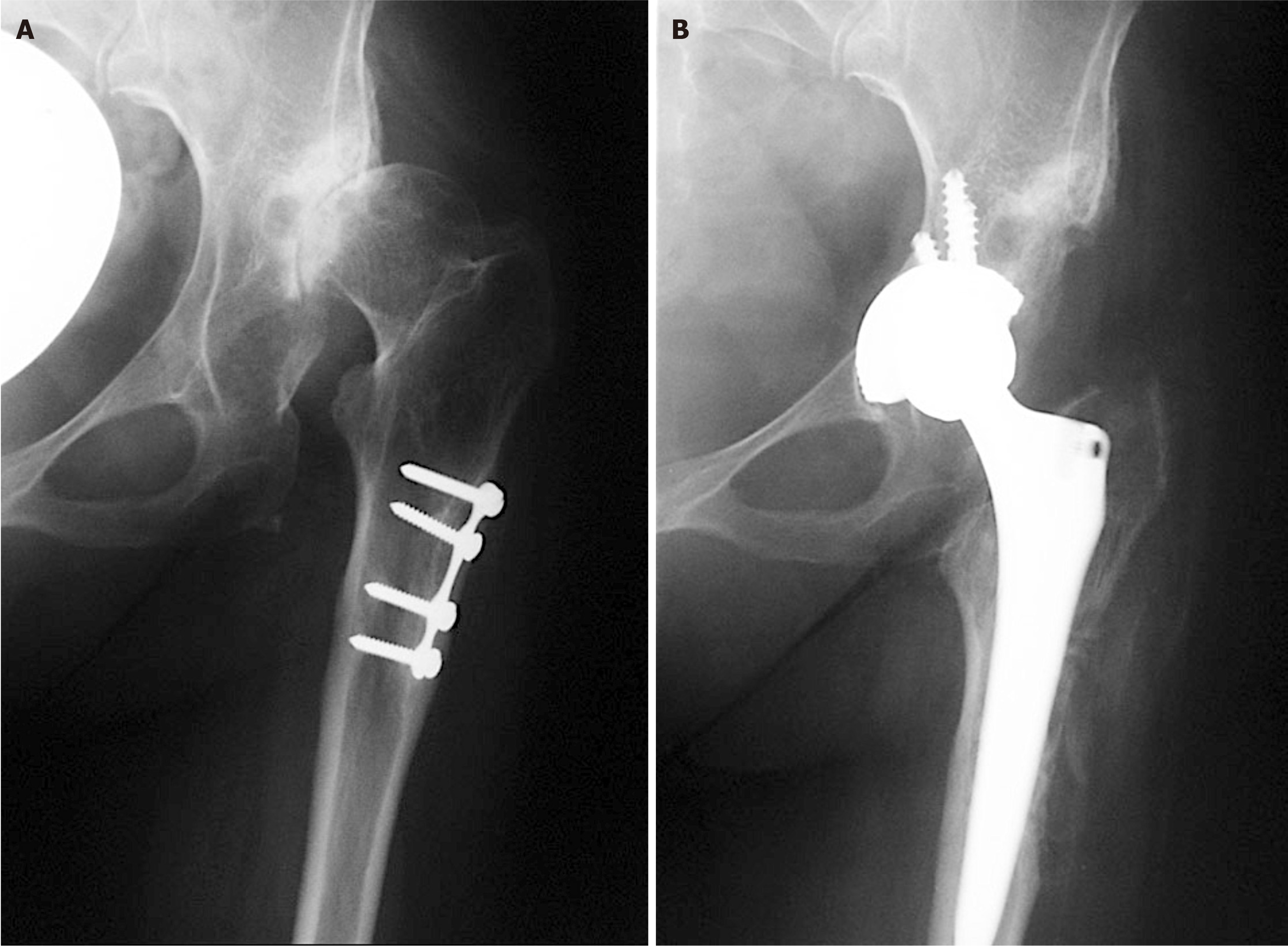
Figure 1 X-rays of left hip.
A: Preoperative X-ray of a patient with secondary osteoarthritis of the left hip due to dysplasia. Plate and screws in the proximal femur are left from the previous surgery performed in childhood; B: Postoperative X-ray with implanted uncemented acetabular cup and femoral stem. Acetabular cup is protruding beyond the Kohler’s line inside the pelvis and secured with 3 additional screws. Lesser trochanter is brought distally to the normal level so there is no leg length discrepancy postoperatively.
- Citation: Barbaric Starcevic K, Bicanic G, Bicanic L. Specific approach to total hip arthroplasty in patients with childhood hip disorders sequelae. World J Orthop 2024; 15(12): 1118-1123
- URL: https://www.wjgnet.com/2218-5836/full/v15/i12/1118.htm
- DOI: https://dx.doi.org/10.5312/wjo.v15.i12.1118


