Copyright
©The Author(s) 2024.
World J Orthop. Nov 18, 2024; 15(11): 1095-1100
Published online Nov 18, 2024. doi: 10.5312/wjo.v15.i11.1095
Published online Nov 18, 2024. doi: 10.5312/wjo.v15.i11.1095
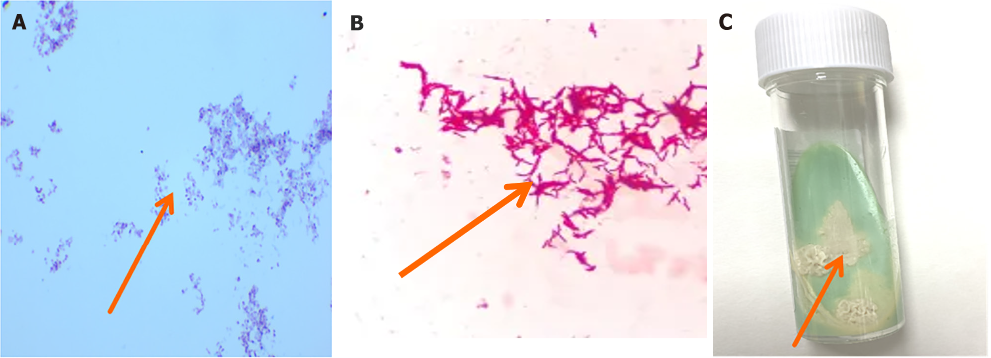
Figure 3 Cultivation of nontuberculous mycobacteria and microscopic images.
A: Positive acid fast staining of pus, with a large number of red elongated hyphae (100 ×); B: Under the microscope, slender, red, rod-shaped nontuberculous mycobacteria could be seen, with relatively scattered distribution and some bacterial cells slightly curved (400 ×); C: On the 12th day of observation, a unique scene appeared on Roche medium. Rice yellowish dry bacterial colonies appeared on top. This rice yellow color was very eye-catching and stood out, particularly against the background of the culture medium. The colony appeared dry and lacked a moist luster.
- Citation: Lin HY, Tan QH. Metagenomic next-generation sequencing may assist diagnosis of osteomyelitis caused by Mycobacterium houstonense: A case report. World J Orthop 2024; 15(11): 1095-1100
- URL: https://www.wjgnet.com/2218-5836/full/v15/i11/1095.htm
- DOI: https://dx.doi.org/10.5312/wjo.v15.i11.1095


