Copyright
©The Author(s) 2024.
World J Orthop. Nov 18, 2024; 15(11): 1056-1074
Published online Nov 18, 2024. doi: 10.5312/wjo.v15.i11.1056
Published online Nov 18, 2024. doi: 10.5312/wjo.v15.i11.1056
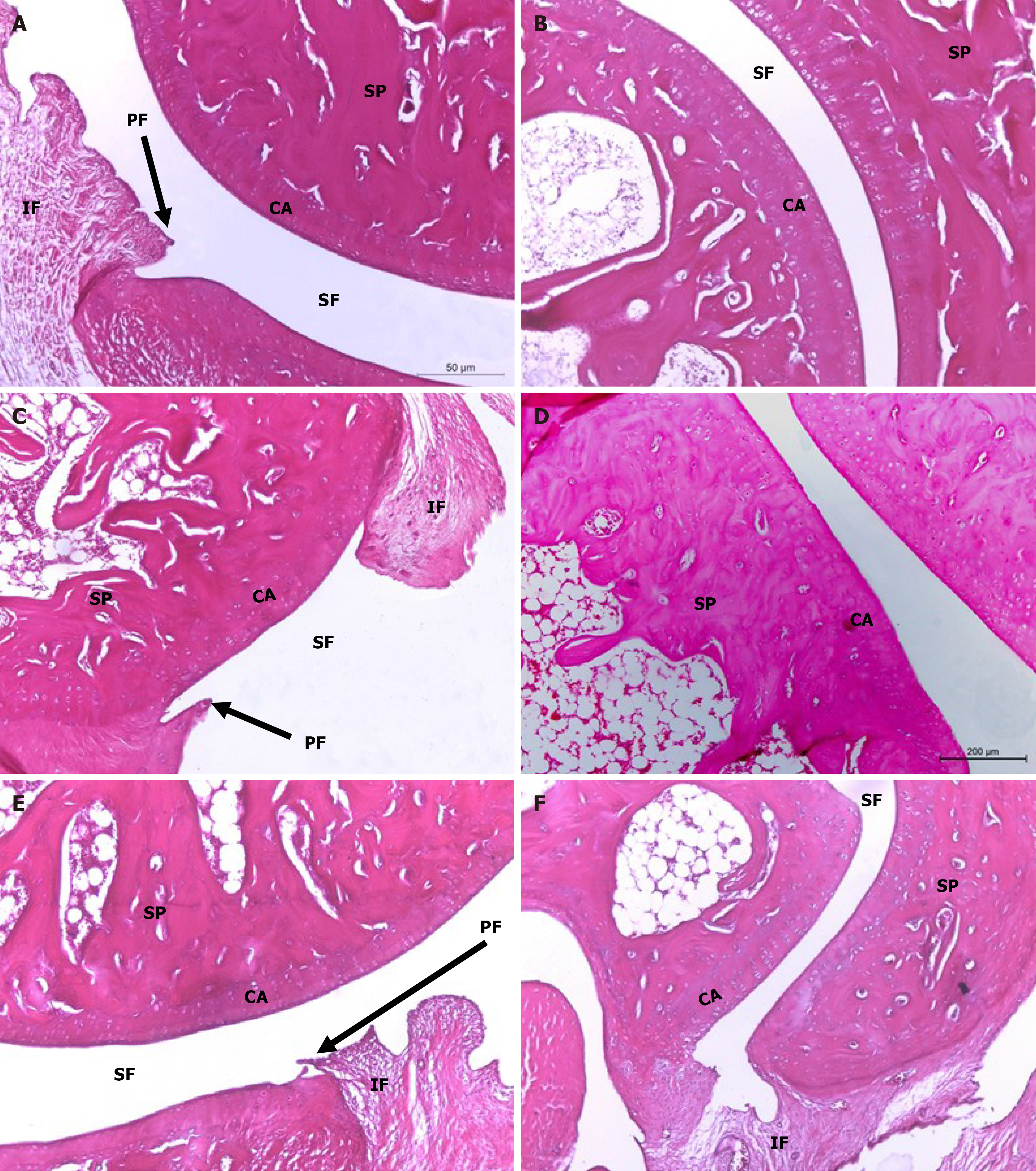
Figure 6 Histopathological evaluation of treatments on monosodium iodate-induced osteoarthritis in rat.
Microphotographs showing the histological changes of hind ankle in hematoxylin and eosin (100 ×) stained sections. A and B: The histological picture of hind ankle joints of osteoarthritic rats treated with hyaluronic acid (HA); C and D: The histological picture of hind ankle joints of osteoarthritic rats treated with bone marrow mesenchymal stem cells (BMMSCs); E and F: Treated with BMMSCs + HA revealed improvements in the ankle joint articular tissue integrity and architecture with suppressed inflammation and reduced synovial membrane thickening. CA: Articular cartilage; EC: Erosion of cartilage; IF: Inflammatory cell infiltration; PF: Pannus formation; SF: Synovial fluid; SM: Synovial membrane; SP: Spongy bone.
- Citation: Hagag UI, Halfaya FM, Al-Muzafar HM, Al-Jameel SS, Amin KA, Abou El-Kheir W, Mahdi EA, Hassan GANR, Ahmed OM. Impacts of mesenchymal stem cells and hyaluronic acid on inflammatory indicators and antioxidant defense in experimental ankle osteoarthritis. World J Orthop 2024; 15(11): 1056-1074
- URL: https://www.wjgnet.com/2218-5836/full/v15/i11/1056.htm
- DOI: https://dx.doi.org/10.5312/wjo.v15.i11.1056


