Copyright
©The Author(s) 2024.
World J Orthop. Oct 18, 2024; 15(10): 981-990
Published online Oct 18, 2024. doi: 10.5312/wjo.v15.i10.981
Published online Oct 18, 2024. doi: 10.5312/wjo.v15.i10.981
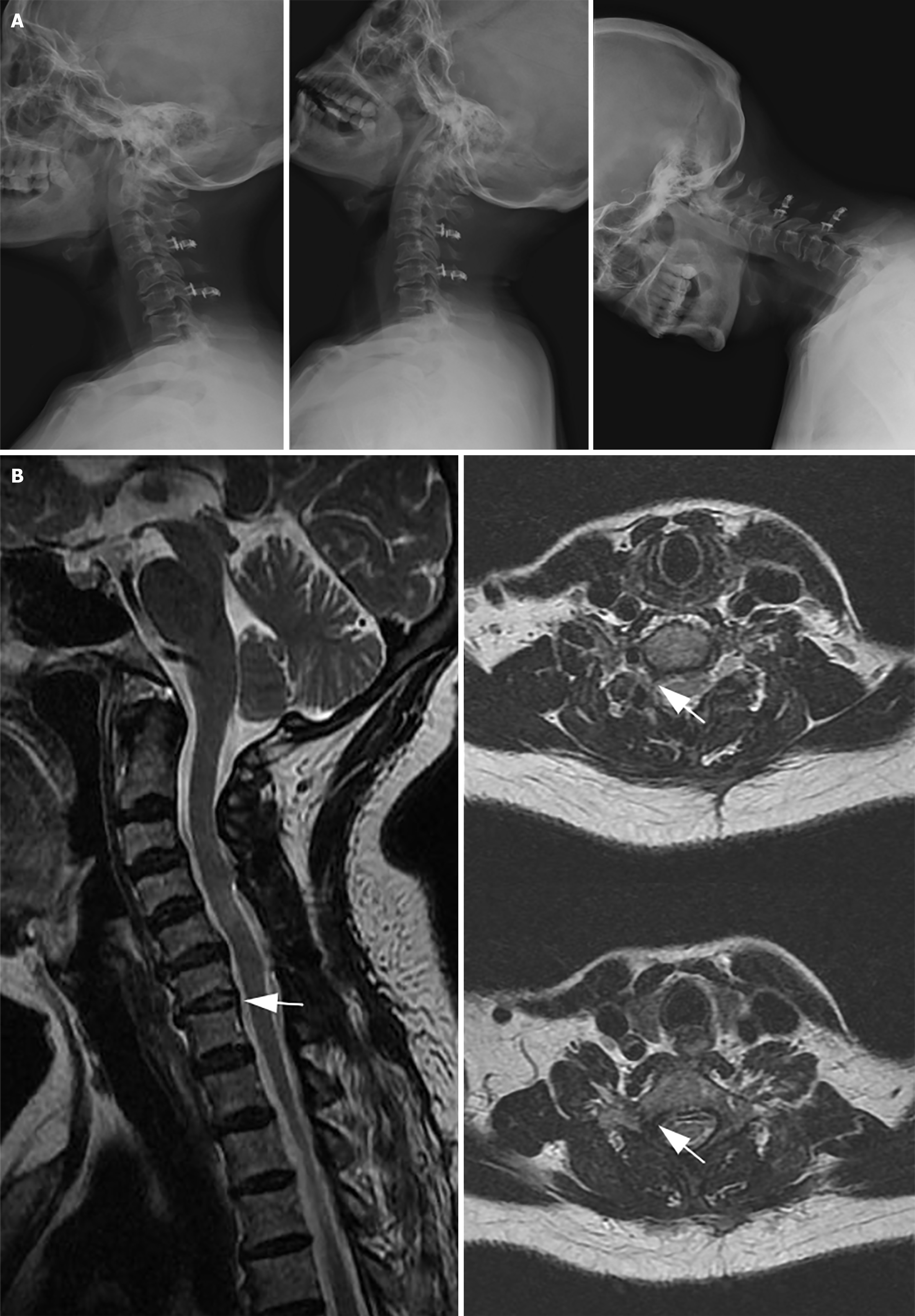
Figure 3 Outpatient follow-up 6 months post-surgery.
A: X-ray examination of the cervical spine in lateral and hyperextension flexion positions displays a significant improvement in the cervical spine's range of motion; B: Magnetic resonance imaging at 6 months post-surgery: The herniated nucleus pulposus tissue of C6/7 has been eliminated, and the right C7 nerve has been entirely decompressed, with no signal from the nucleus pulposus tissue (indicated by the white arrow).
- Citation: Cui HC, Chang ZQ, Zhao SK. Atypical cervical spondylotic radiculopathy resulting in a hypertensive emergency during cervical extension: A case report and review of literature. World J Orthop 2024; 15(10): 981-990
- URL: https://www.wjgnet.com/2218-5836/full/v15/i10/981.htm
- DOI: https://dx.doi.org/10.5312/wjo.v15.i10.981


