Copyright
©The Author(s) 2024.
World J Orthop. Oct 18, 2024; 15(10): 981-990
Published online Oct 18, 2024. doi: 10.5312/wjo.v15.i10.981
Published online Oct 18, 2024. doi: 10.5312/wjo.v15.i10.981
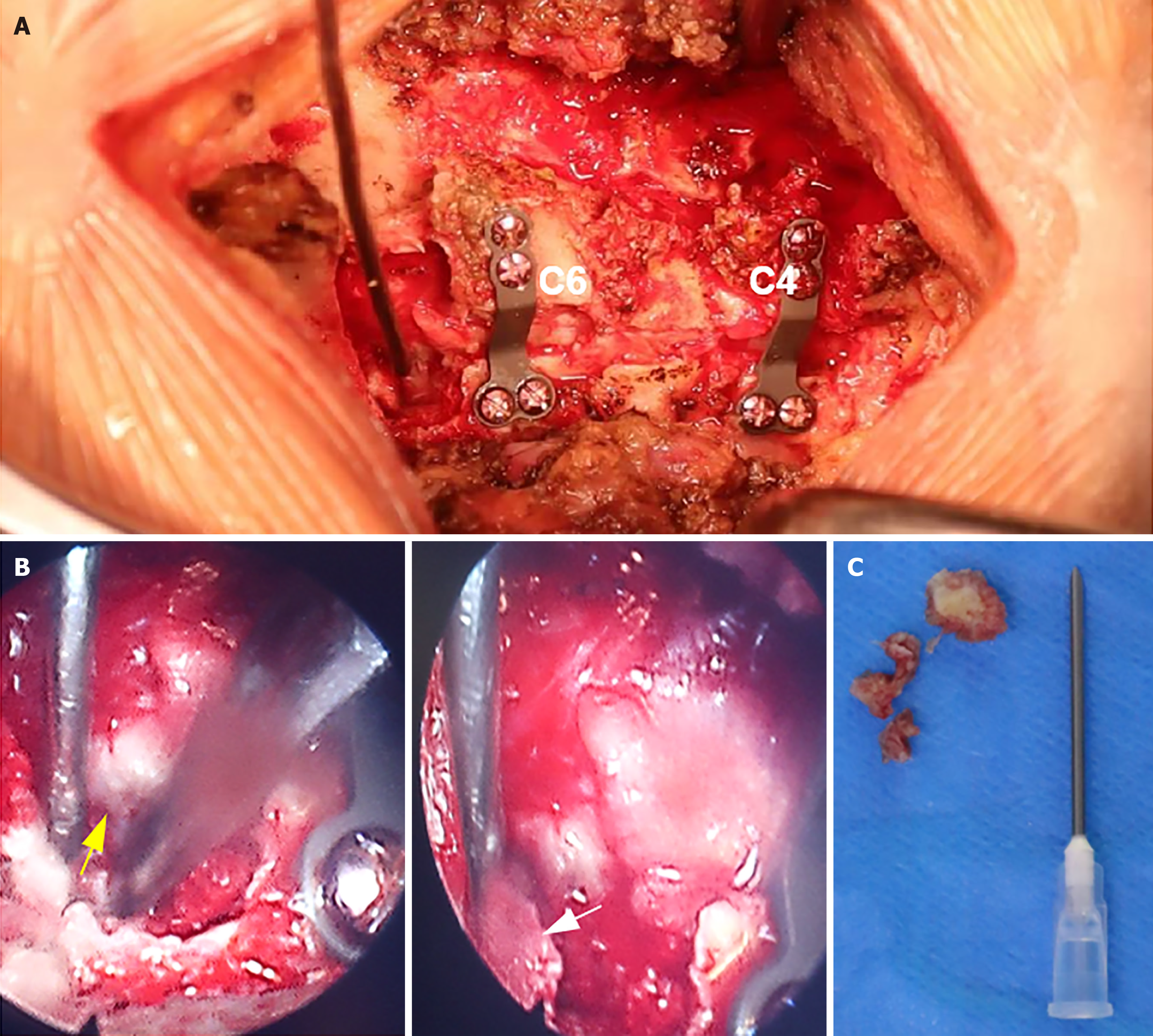
Figure 2 Intraoperative image.
A: The vertebral laminae of C4 and C6 were elevated, facilitating decompression of the spinal cord, followed by placement of an internal fixation plate; B: The ventral nucleus pulposus was extracted from the right nerve root of C7 using foraminal mirror-assisted extraction technique. During this procedure, the dural membrane (indicated by the yellow arrow) and excised free nucleus pulposus tissue (indicated by the white arrow) were visually observed; C: Intraoperatively obtained free nucleus pulposus tissue from the ventral aspect of the C7 nerve root.
- Citation: Cui HC, Chang ZQ, Zhao SK. Atypical cervical spondylotic radiculopathy resulting in a hypertensive emergency during cervical extension: A case report and review of literature. World J Orthop 2024; 15(10): 981-990
- URL: https://www.wjgnet.com/2218-5836/full/v15/i10/981.htm
- DOI: https://dx.doi.org/10.5312/wjo.v15.i10.981


