Copyright
©The Author(s) 2024.
World J Orthop. Oct 18, 2024; 15(10): 981-990
Published online Oct 18, 2024. doi: 10.5312/wjo.v15.i10.981
Published online Oct 18, 2024. doi: 10.5312/wjo.v15.i10.981
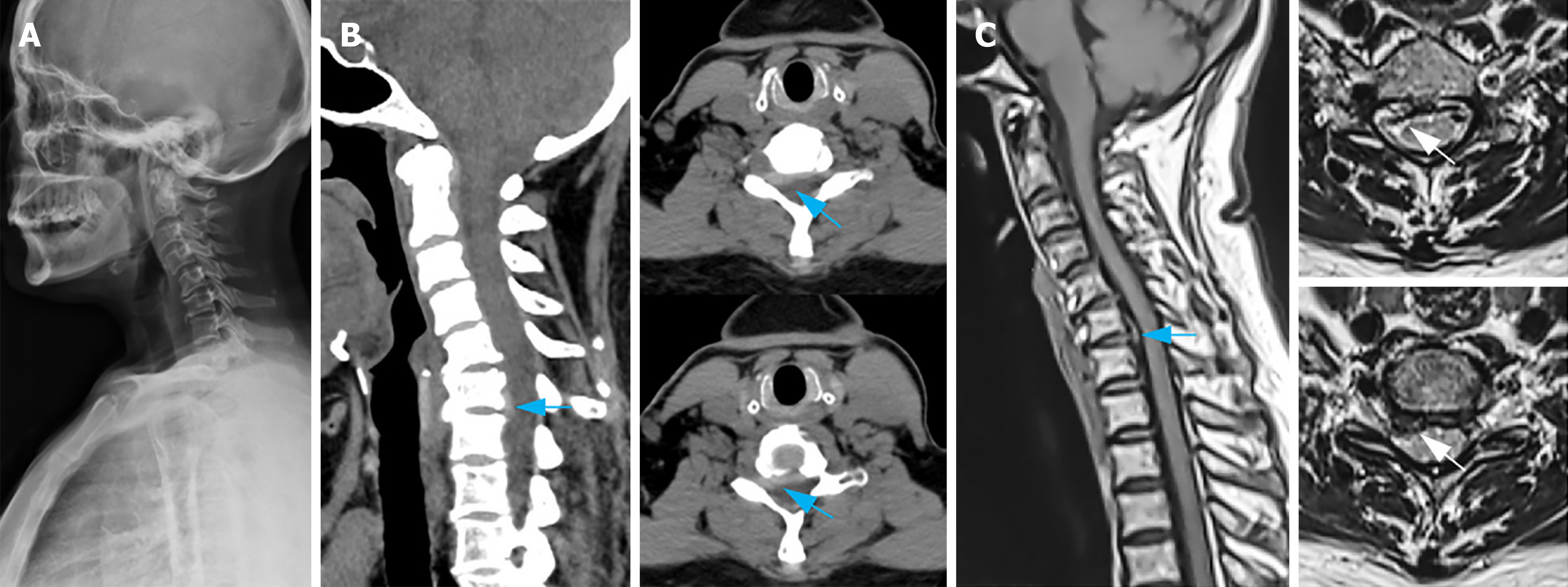
Figure 1 Preoperative imaging examinations.
A: A lateral plain film of the cervical vertebra depicts cervical vertebra's reversed arch, narrowed C6/7 space, and patchy calcification of the nuchal ligament; B: Cervical vertebra computerized tomography sagittal reconstruction and plain scan demonstrate an intervertebral disc prolapse and upward displacement in the C6/7 space, along with right nerve root canal stenosis (indicated by the blue arrow); C: Sagittal magnetic resonance imaging and plain scan of cervical vertebra present herniation and upward displacement of the intervertebral disc in the C6/7 space (indicated by the blue arrow), discontinuous intervertebral disc signal, and compression of the right C7 nerve root (indicated by the white arrow).
- Citation: Cui HC, Chang ZQ, Zhao SK. Atypical cervical spondylotic radiculopathy resulting in a hypertensive emergency during cervical extension: A case report and review of literature. World J Orthop 2024; 15(10): 981-990
- URL: https://www.wjgnet.com/2218-5836/full/v15/i10/981.htm
- DOI: https://dx.doi.org/10.5312/wjo.v15.i10.981


