Copyright
©The Author(s) 2024.
World J Orthop. Oct 18, 2024; 15(10): 973-980
Published online Oct 18, 2024. doi: 10.5312/wjo.v15.i10.973
Published online Oct 18, 2024. doi: 10.5312/wjo.v15.i10.973
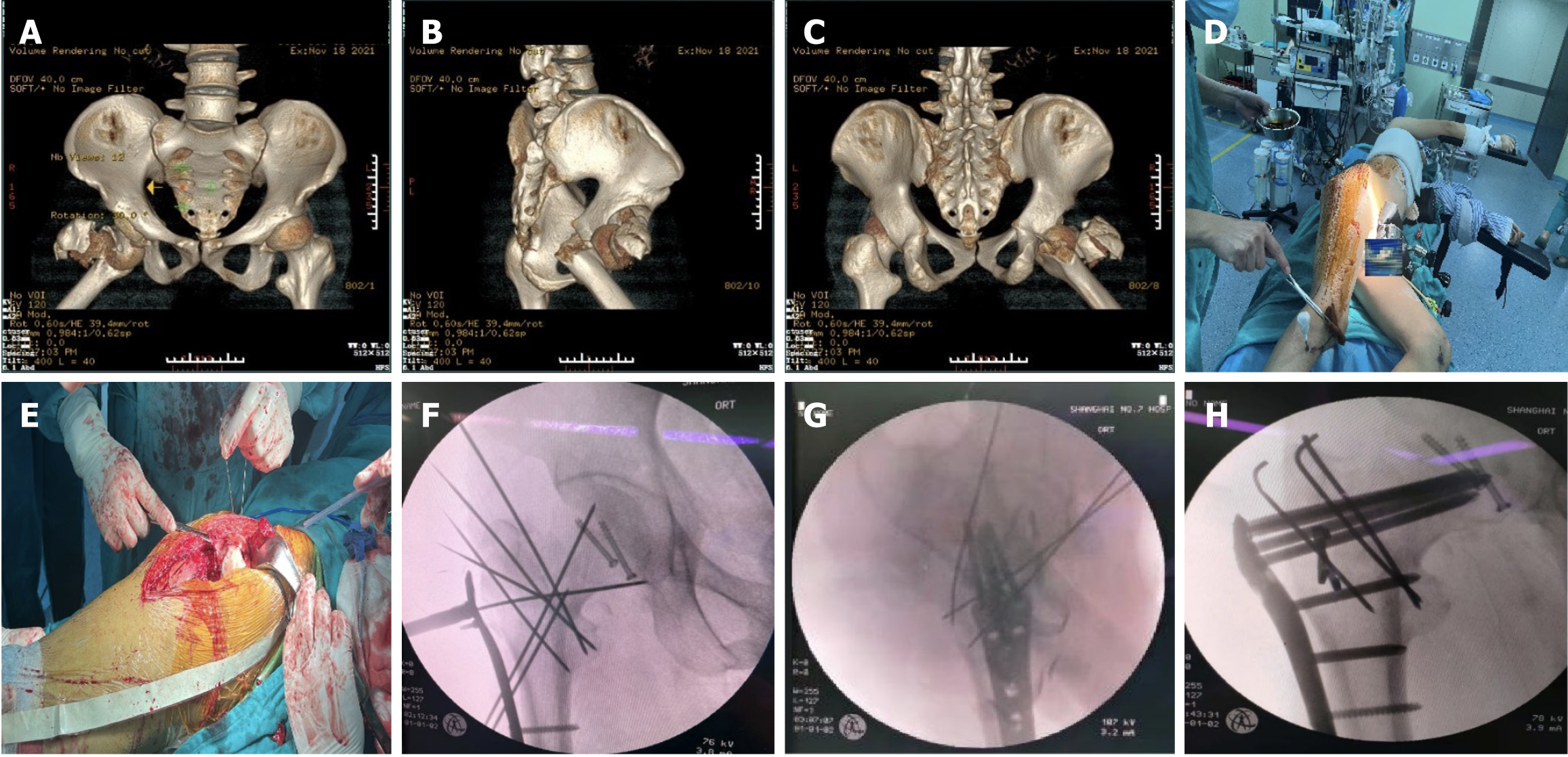
Figure 2 Preoperative and intraoperative images.
A: Front view of preoperative 3-dimensional (3D) computed tomography (CT); B: Lateral view of preoperative 3D CT; C: Back view of preoperative 3D CT. 3D CT revealed that the greater trochanter was fractured and that the lesser trochanter was intact, belonging to AO/OTA A1.1; D: Surgical position; E: Surgical incision; F: Intraoperative Kirschner wire temporary fixation; G: Lateral radiograph from intraoperative fluoroscopy; H: Anteroposterior radiograph from intraoperative fluoroscopy.
- Citation: Yu X, Li YZ, Lu HJ, Liu BL. Treatment of a femoral neck fracture combined with ipsilateral femoral head and intertrochanteric fractures: A case report. World J Orthop 2024; 15(10): 973-980
- URL: https://www.wjgnet.com/2218-5836/full/v15/i10/973.htm
- DOI: https://dx.doi.org/10.5312/wjo.v15.i10.973


