Copyright
©The Author(s) 2024.
World J Orthop. Oct 18, 2024; 15(10): 965-972
Published online Oct 18, 2024. doi: 10.5312/wjo.v15.i10.965
Published online Oct 18, 2024. doi: 10.5312/wjo.v15.i10.965
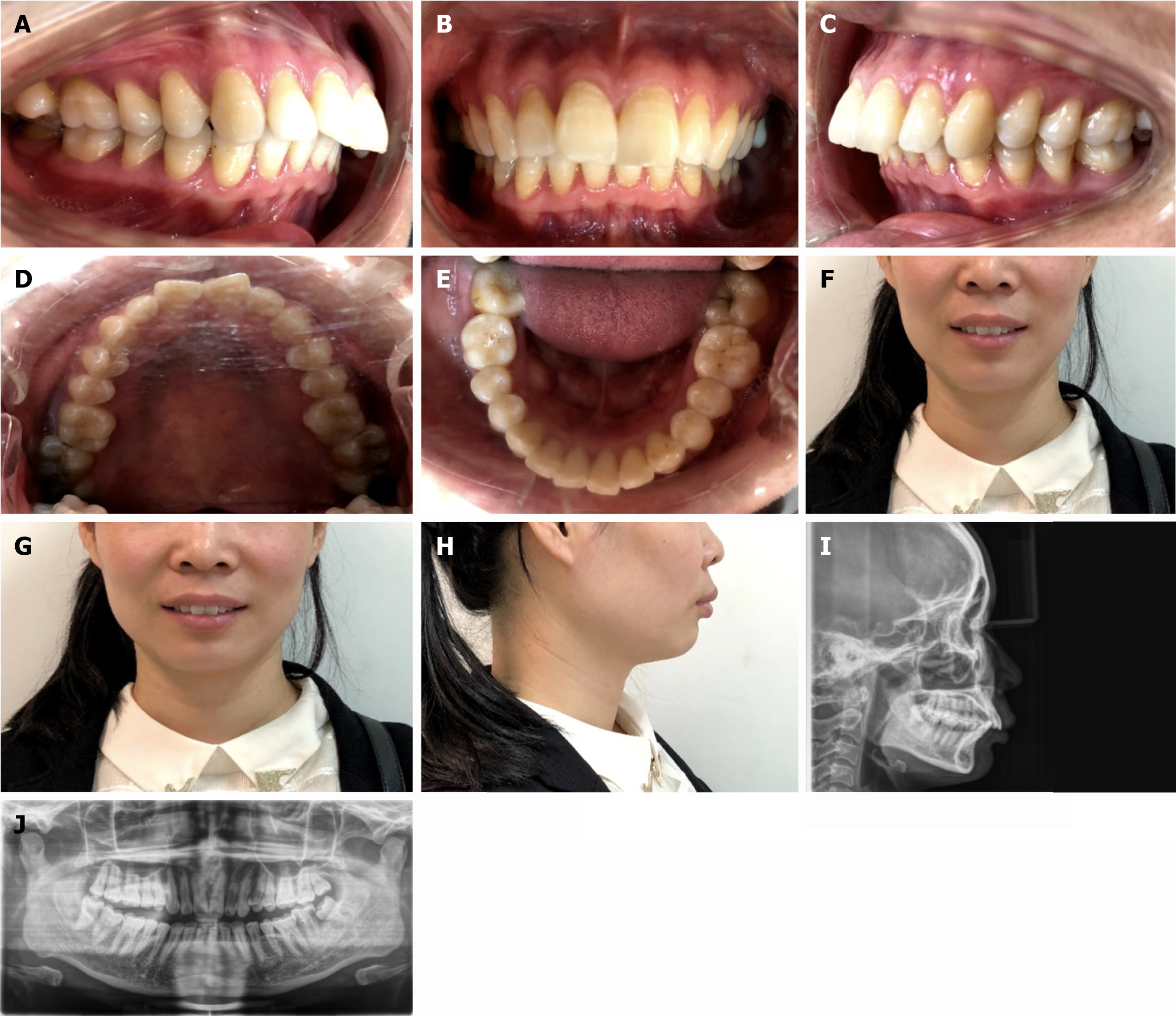
Figure 1 Pretreatment intraoral and facial photographs and panoramic radiographs.
A: The right lateral view and the scissors bite of the second permanent molars; B: The frontal view and deep overjet; C: The left lateral view and the scissors bite of the second permanent molar; D: The palatal view; E: The lingual view; F: Frontal image; G: Smiling image; H: Profile image; I: Cephalometric; J: Panoramic radiograph.
- Citation: Xie LL, Chu DY, Wu XF. Simple and effective method for treating severe adult skeletal class II malocclusion: A case report. World J Orthop 2024; 15(10): 965-972
- URL: https://www.wjgnet.com/2218-5836/full/v15/i10/965.htm
- DOI: https://dx.doi.org/10.5312/wjo.v15.i10.965


