Copyright
©The Author(s) 2023.
World J Orthop. May 18, 2023; 14(5): 362-368
Published online May 18, 2023. doi: 10.5312/wjo.v14.i5.362
Published online May 18, 2023. doi: 10.5312/wjo.v14.i5.362
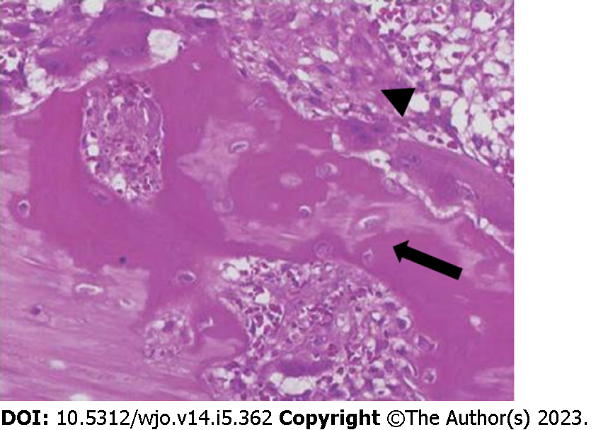
Figure 3 Microscopic findings of myositis ossificans.
Haematoxylin and eosin staining showing a mature lamellar bone neo-formation (arrow) and fibroblasts with elongated nuclei (arrowhead) arranged in short irregular fascicles with the typical "zonation pattern" placed in loose myxoid or collagenous stroma. Occasional mitoses can be observed, in the absence of cellular atypia.
- Citation: Carbone G, Andreasi V, De Nardi P. Intra-abdominal myositis ossificans - a clinically challenging disease: A case report. World J Orthop 2023; 14(5): 362-368
- URL: https://www.wjgnet.com/2218-5836/full/v14/i5/362.htm
- DOI: https://dx.doi.org/10.5312/wjo.v14.i5.362


