Copyright
©The Author(s) 2023.
World J Orthop. May 18, 2023; 14(5): 362-368
Published online May 18, 2023. doi: 10.5312/wjo.v14.i5.362
Published online May 18, 2023. doi: 10.5312/wjo.v14.i5.362
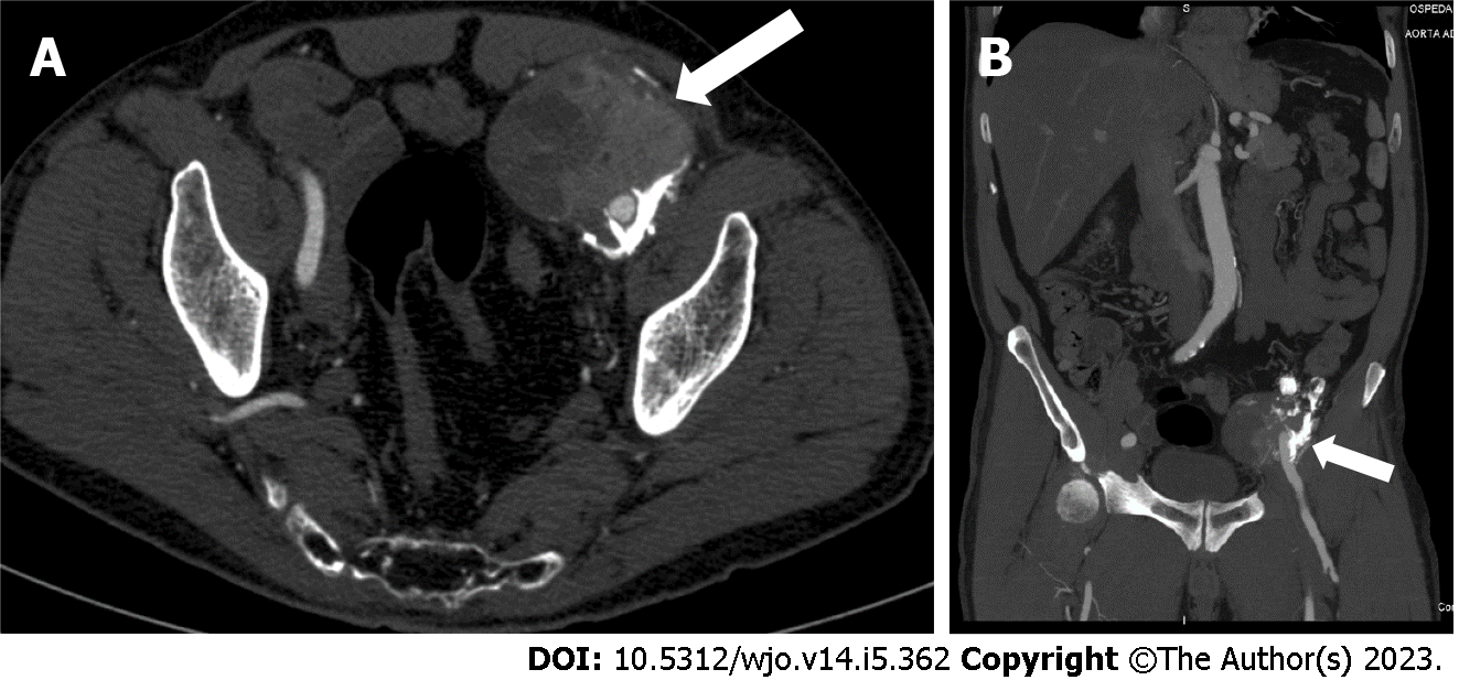
Figure 2 Computed tomography image revealed an 8 cm × 6.
5 cm × 7 cm, highly inhomogeneous mass composed by multiple calcifications at the periphery. A: Axial view; B: Sagittal view.
- Citation: Carbone G, Andreasi V, De Nardi P. Intra-abdominal myositis ossificans - a clinically challenging disease: A case report. World J Orthop 2023; 14(5): 362-368
- URL: https://www.wjgnet.com/2218-5836/full/v14/i5/362.htm
- DOI: https://dx.doi.org/10.5312/wjo.v14.i5.362


