Copyright
©The Author(s) 2023.
World J Orthop. May 18, 2023; 14(5): 362-368
Published online May 18, 2023. doi: 10.5312/wjo.v14.i5.362
Published online May 18, 2023. doi: 10.5312/wjo.v14.i5.362
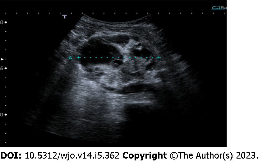
Figure 1 Abdominal ultrasound showed a 7-cm ovoidal lesion with a mixed echo pattern.
It was possible to distinguish fluid and solid components (including diffuse calcifications).
- Citation: Carbone G, Andreasi V, De Nardi P. Intra-abdominal myositis ossificans - a clinically challenging disease: A case report. World J Orthop 2023; 14(5): 362-368
- URL: https://www.wjgnet.com/2218-5836/full/v14/i5/362.htm
- DOI: https://dx.doi.org/10.5312/wjo.v14.i5.362


