Copyright
©The Author(s) 2023.
World J Orthop. Apr 18, 2023; 14(4): 171-185
Published online Apr 18, 2023. doi: 10.5312/wjo.v14.i4.171
Published online Apr 18, 2023. doi: 10.5312/wjo.v14.i4.171
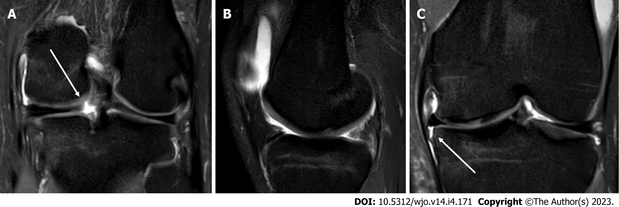
Figure 8 Isolated posterior root tear of the medial meniscus.
A: T2 Fat-Sat coronal magnetic resonance imaging (MRI) view, showing disruption of the meniscus ring (white arrow); B: T2 Fat-Sat sagittal MRI view, with the “ghost sign”, highly suggestive for posterior root tear; C: T2 Fat-Sat coronal MRI view, showing detachment of the medial meniscotibial ligament (white arrow) and meniscus extrusion.
- Citation: Simonetta R, Russo A, Palco M, Costa GG, Mariani PP. Meniscus tears treatment: The good, the bad and the ugly-patterns classification and practical guide. World J Orthop 2023; 14(4): 171-185
- URL: https://www.wjgnet.com/2218-5836/full/v14/i4/171.htm
- DOI: https://dx.doi.org/10.5312/wjo.v14.i4.171


