Copyright
©The Author(s) 2023.
World J Orthop. Mar 18, 2023; 14(3): 136-145
Published online Mar 18, 2023. doi: 10.5312/wjo.v14.i3.136
Published online Mar 18, 2023. doi: 10.5312/wjo.v14.i3.136
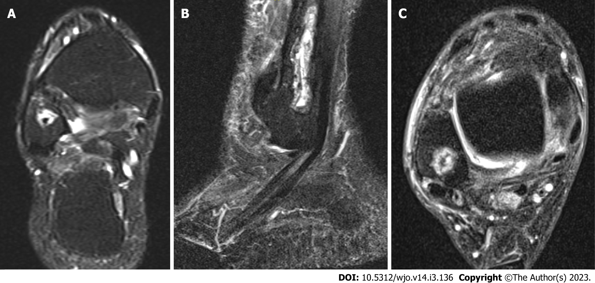
Figure 3 Ankle magnetic resonance imaging of case 2 showing a Broadie’s abscess.
A: Coronal; B: Sagittal; C: Axial T2-magnetic resonance imaging images showing a hyperintense intra-osseous collection consistent with Broadie’s abscess. The patient did not show any elevated inflammatory markers throughout the treatment.
- Citation: Ahmed AH, Ahmed S, Barakat A, Mangwani J, White H. Inflammatory response in confirmed non-diabetic foot and ankle infections: A case series with normal inflammatory markers. World J Orthop 2023; 14(3): 136-145
- URL: https://www.wjgnet.com/2218-5836/full/v14/i3/136.htm
- DOI: https://dx.doi.org/10.5312/wjo.v14.i3.136


