Copyright
©The Author(s) 2023.
World J Orthop. Dec 18, 2023; 14(12): 868-877
Published online Dec 18, 2023. doi: 10.5312/wjo.v14.i12.868
Published online Dec 18, 2023. doi: 10.5312/wjo.v14.i12.868
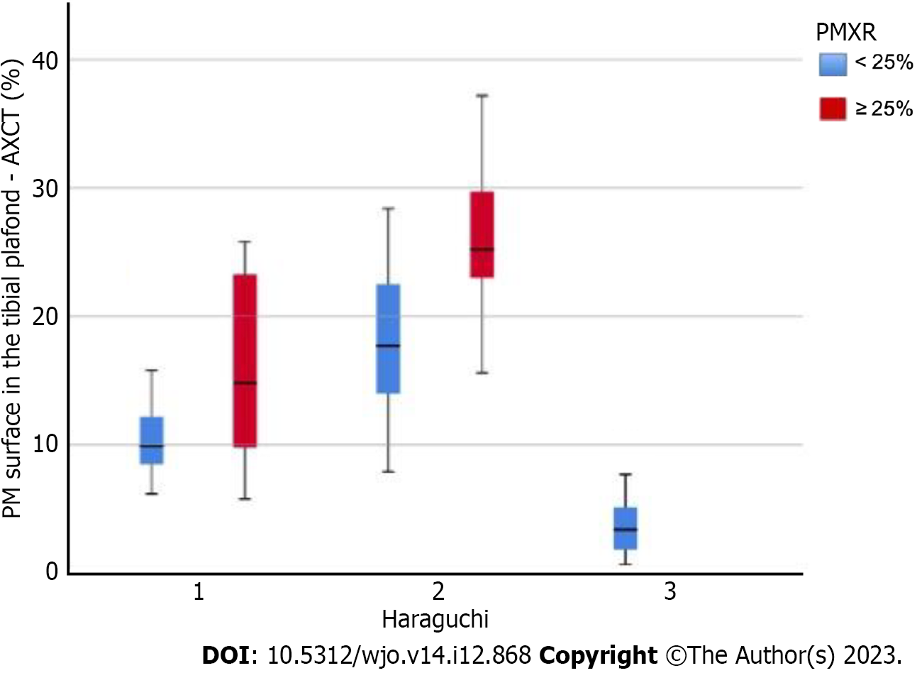
Figure 5 The distribution of the percent area of the tibial plafond of the posterior malleolus fracture according to the various Haraguchi classification types.
Blue: Group A; Red: Group B; PMXR: Posterior malleolus size in profile X-ray images; AXCT: Axial computed tomography slice of the posterior malleolus; PM: Posterior malleolus.
- Citation: De Marchi Neto N, Nesello PFT, Bergamasco JM, Costa MT, Christian RW, Severino NR. Importance of computed tomography in posterior malleolar fractures: Added information to preoperative X-ray studies. World J Orthop 2023; 14(12): 868-877
- URL: https://www.wjgnet.com/2218-5836/full/v14/i12/868.htm
- DOI: https://dx.doi.org/10.5312/wjo.v14.i12.868


