Copyright
©The Author(s) 2023.
World J Orthop. Nov 18, 2023; 14(11): 827-835
Published online Nov 18, 2023. doi: 10.5312/wjo.v14.i11.827
Published online Nov 18, 2023. doi: 10.5312/wjo.v14.i11.827
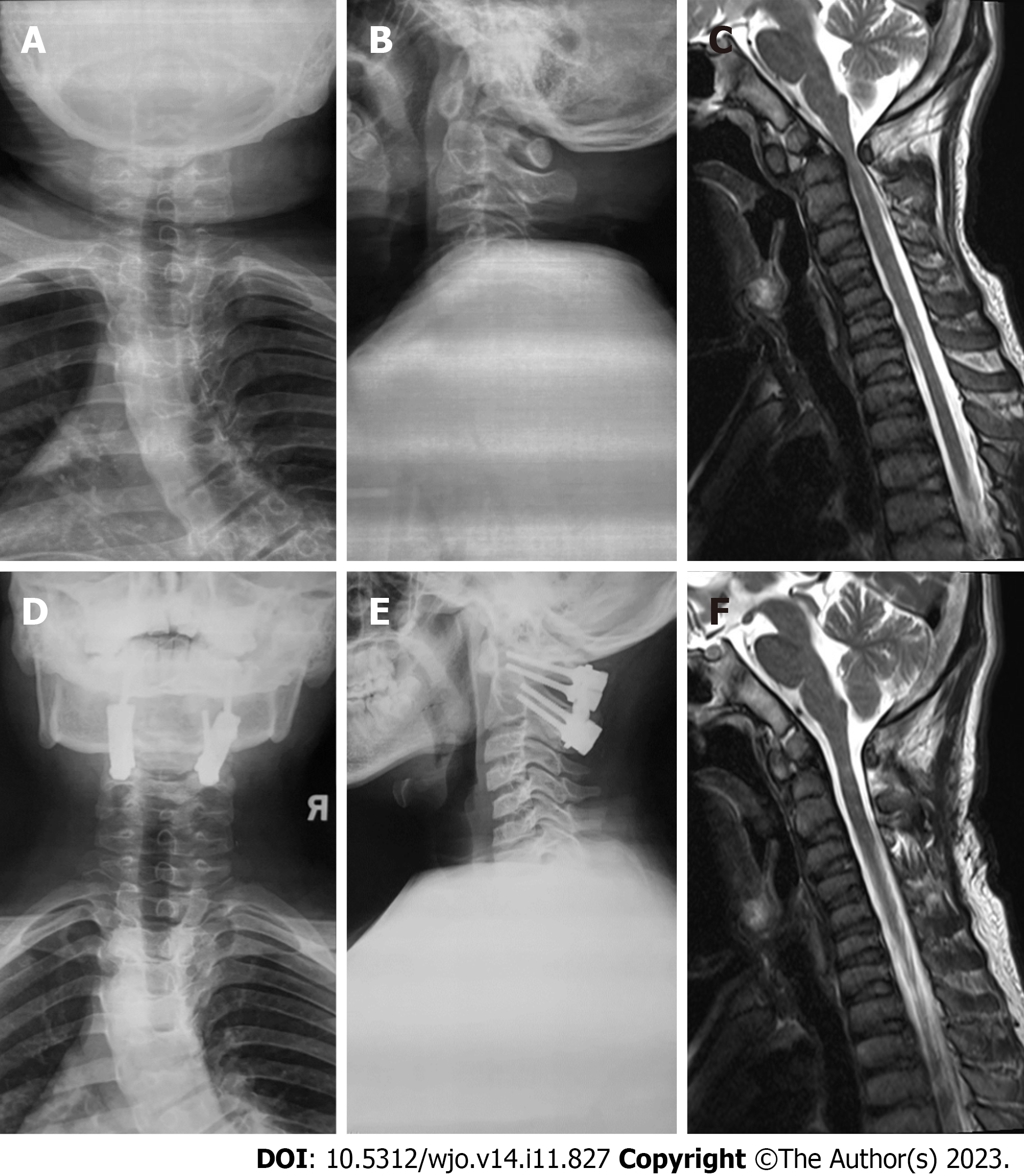
Figure 4 Imaging data of atlantoaxial dysplasia before and after surgical treatment.
A and B: Preoperative X-ray shows the widening of space between the anterior arch and odontoid process and dysplasia of the odontoid process; C: Preoperative MRI shows atlas-level spinal canal stenosis, spinal cord compression, and mild degeneration; D and E: Postoperative X-ray shows atlantoaxial fixation and fusion; F: The compression of the atlantoaxial spinal cord is relieved after the operation.
- Citation: Jiao Y, Zhao JD, Huang XA, Cai HY, Shen JX. Surgical treatment of atlantoaxial dysplasia and scoliosis in spondyloepiphyseal dysplasia congenita: A case report. World J Orthop 2023; 14(11): 827-835
- URL: https://www.wjgnet.com/2218-5836/full/v14/i11/827.htm
- DOI: https://dx.doi.org/10.5312/wjo.v14.i11.827


