Copyright
©The Author(s) 2023.
World J Orthop. Nov 18, 2023; 14(11): 800-812
Published online Nov 18, 2023. doi: 10.5312/wjo.v14.i11.800
Published online Nov 18, 2023. doi: 10.5312/wjo.v14.i11.800
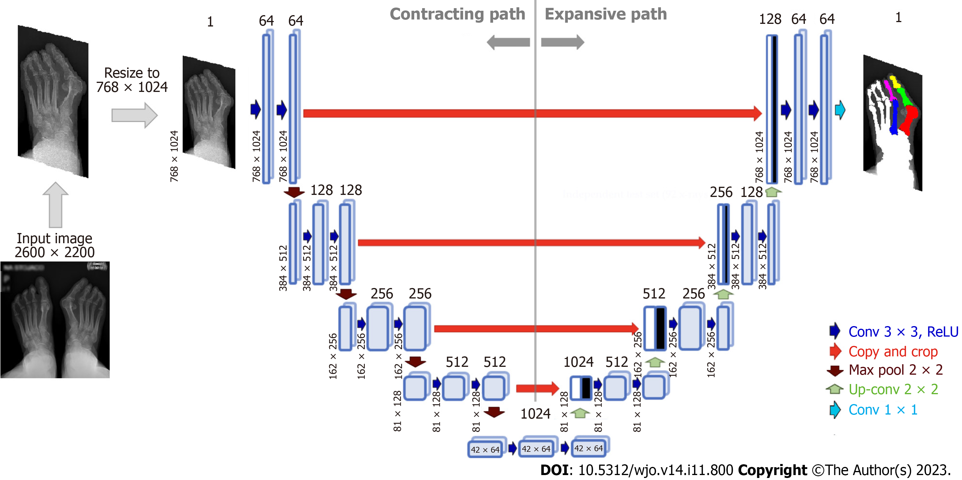
Figure 4 Architecture of U-Net neural network for bone segmentation from radiographs.
The network consists of an encoder (contracting path), which encodes an input image size of 768 × 1024 pixels to 42 × 64 × 1024 feature tensor at the bottleneck, and a decoder (expanding path) that decodes the feature tensor to the segmented image of the same size as the input image. The decoder follows the typical architecture of a convolutional network. Each block in the decoder consists of two 3 × 3 convolutions, each followed by a rectified linear unit (ReLU) and a 2 × 2 max pooling with stride 2 for down-sampling. Each down-sampling step of the encoder doubles the number of feature channels and decreases the image resolution by half. Every block in the decoder comprises upsampling the feature map followed by a 2 × 2 convolution that halves the number of feature channels - a concatenation with the correspondingly cropped feature map from the contracting path - and two 3 × 3 convolutions, each followed by a ReLU.
- Citation: Kwolek K, Gądek A, Kwolek K, Kolecki R, Liszka H. Automated decision support for Hallux Valgus treatment options using anteroposterior foot radiographs. World J Orthop 2023; 14(11): 800-812
- URL: https://www.wjgnet.com/2218-5836/full/v14/i11/800.htm
- DOI: https://dx.doi.org/10.5312/wjo.v14.i11.800


