Copyright
©The Author(s) 2022.
World J Orthop. Feb 18, 2022; 13(2): 178-192
Published online Feb 18, 2022. doi: 10.5312/wjo.v13.i2.178
Published online Feb 18, 2022. doi: 10.5312/wjo.v13.i2.178
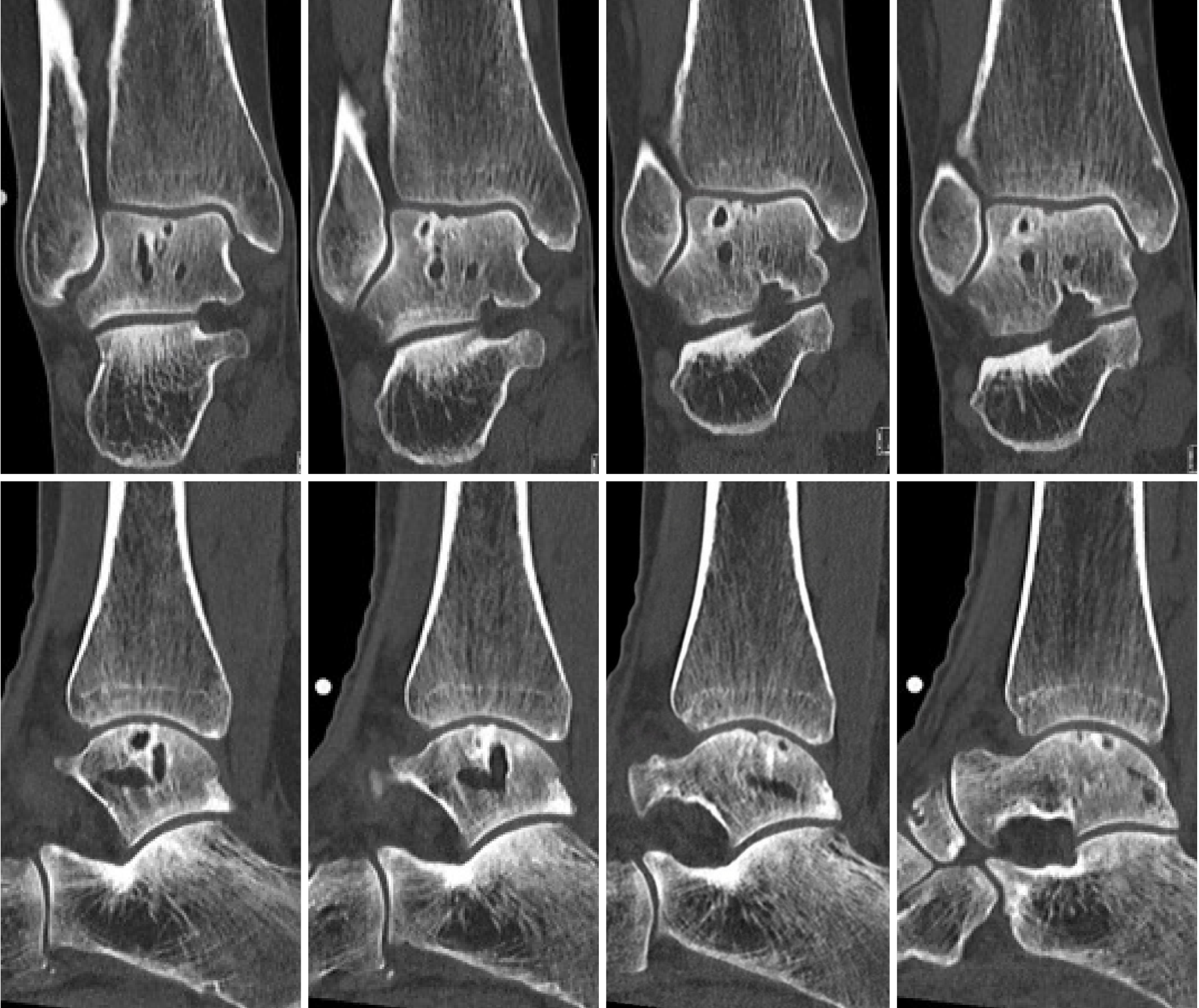
Figure 6 Pre-operative computed tomography scan of patient number 2: Upper part shows coronal slides with the images from left to right going into the posterior to anterior direction.
The lower part shows sagittal slides with the images from left to right going from lateral to medial.
- Citation: Dahmen J, Altink JN, Vuurberg G, Wijdicks CA, Stufkens SA, Kerkhoffs GM. Clinical efficacy of the Ankle Spacer for the treatment of multiple secondary osteochondral lesions of the talus. World J Orthop 2022; 13(2): 178-192
- URL: https://www.wjgnet.com/2218-5836/full/v13/i2/178.htm
- DOI: https://dx.doi.org/10.5312/wjo.v13.i2.178


