Copyright
©The Author(s) 2022.
World J Orthop. Feb 18, 2022; 13(2): 122-130
Published online Feb 18, 2022. doi: 10.5312/wjo.v13.i2.122
Published online Feb 18, 2022. doi: 10.5312/wjo.v13.i2.122
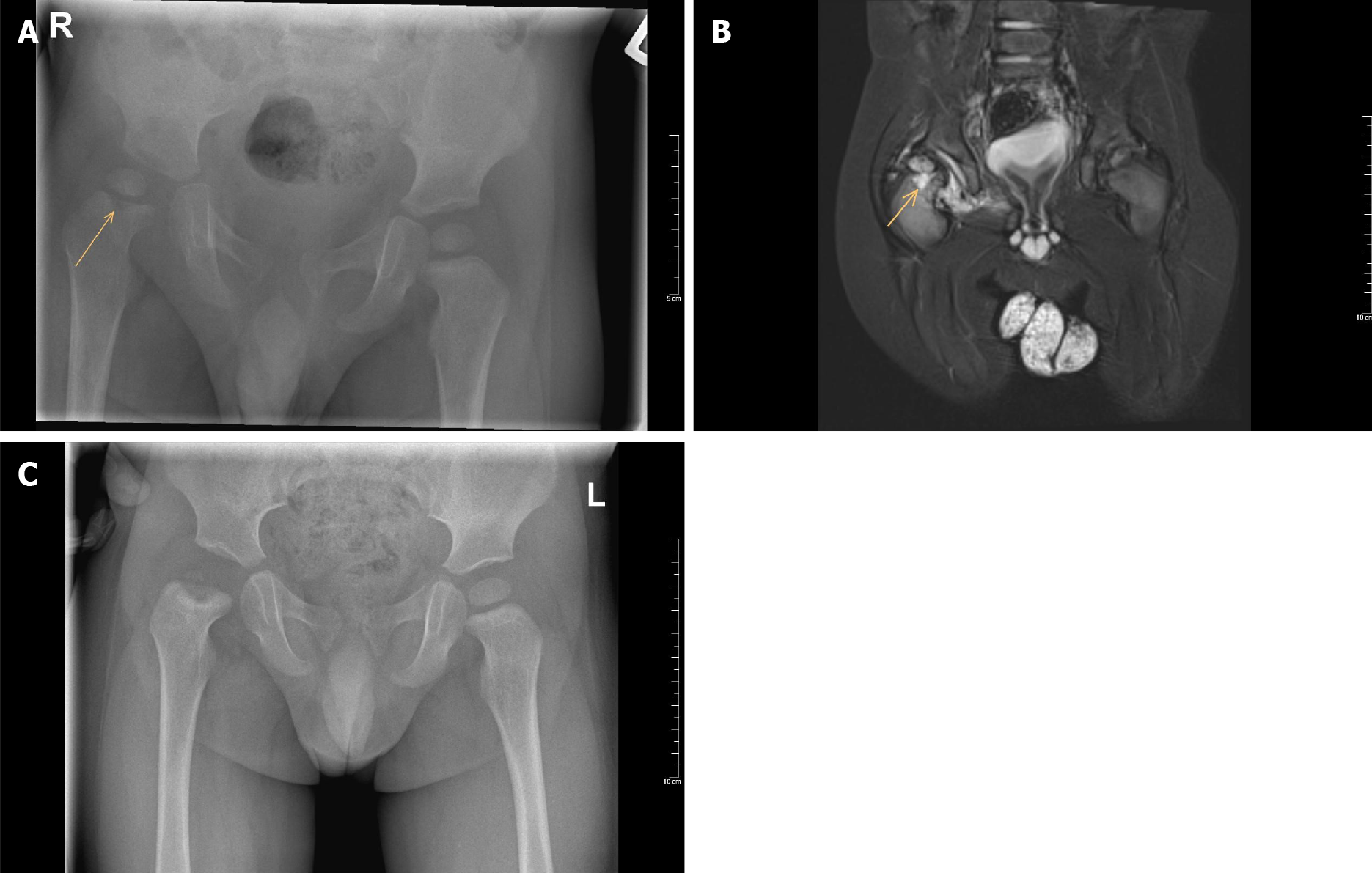
Figure 1 Radiograph images.
A: Plain anteroposterior pelvic radiograph of a one-year-old boy with septic hip arthritis showing concomitant osteomyelitis of the proximal femur at the right side (arrow); B: T2 magnetic resonance imaging coronal view confirms joint effusion, suggestive of hip arthritis, and increased signal of the metaphysis, suggestive of osteomyelitis (arrow); C: Plain anteroposterior radiograph of the same boy after six months follow-up, which shows avascular necrosis of the femoral head.
- Citation: Donders CM, Spaans AJ, van Wering H, van Bergen CJ. Developments in diagnosis and treatment of paediatric septic arthritis. World J Orthop 2022; 13(2): 122-130
- URL: https://www.wjgnet.com/2218-5836/full/v13/i2/122.htm
- DOI: https://dx.doi.org/10.5312/wjo.v13.i2.122


