Copyright
©The Author(s) 2022.
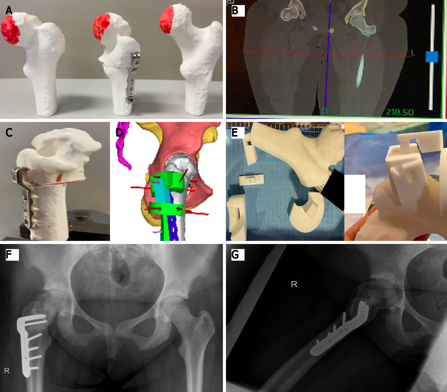
Figure 3 Three-dimensional printing for grade 3 slipped capital femoral epiphysis.
This figure shows different steps required in a case of grade 3 slipped capital femoral epiphysis where three-dimensional (3D)-based templates for positioning of the implant were used, and guidance of the osteotomy during the surgical procedure was performed. A: Preoperative 3D-printed model of the deformed femoral head; B: High-resolution computed tomography scan for exact preoperative planning of deformity correction; C: Preoperative 3D-printed model of the deformed femoral head after correction; D: Analysis of the unique blade plate through 3D-computed view; E: The 3D-printed unique locking system; F: Postoperative anteroposterior radiograph; G: Postoperative lateral radiograph.
- Citation: Goetstouwers S, Kempink D, The B, Eygendaal D, van Oirschot B, van Bergen CJ. Three-dimensional printing in paediatric orthopaedic surgery. World J Orthop 2022; 13(1): 1-10
- URL: https://www.wjgnet.com/2218-5836/full/v13/i1/1.htm
- DOI: https://dx.doi.org/10.5312/wjo.v13.i1.1


