Copyright
©The Author(s) 2021.
World J Orthop. Sep 18, 2021; 12(9): 720-726
Published online Sep 18, 2021. doi: 10.5312/wjo.v12.i9.720
Published online Sep 18, 2021. doi: 10.5312/wjo.v12.i9.720
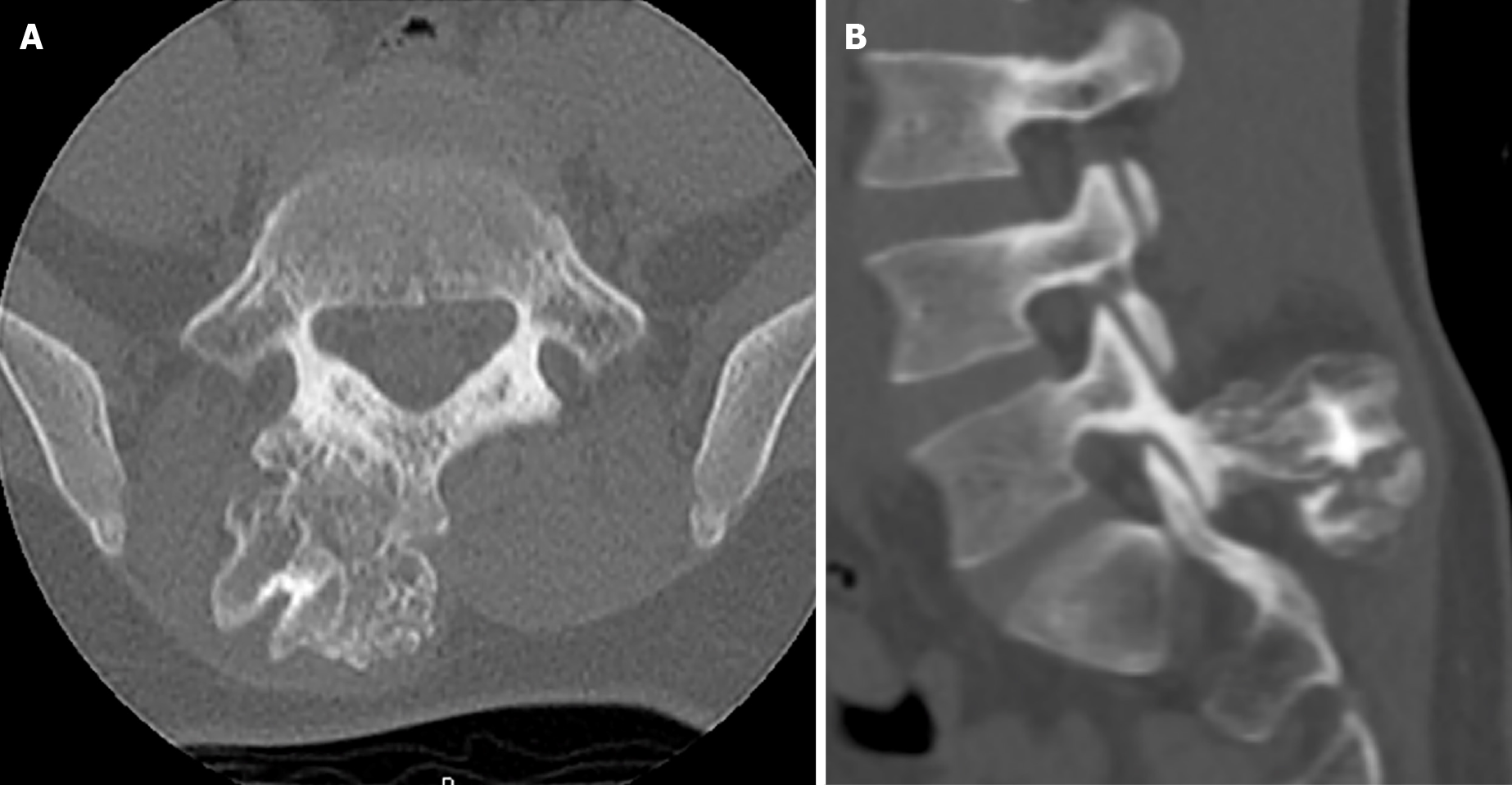
Figure 2 Computed tomography imagines.
A: Axial computed tomography imaging demonstrating osteochondroma protruding from right spinous process/Lamina junction; B: Sagittal computed tomography imaging at the level of the L5/S1 facet joint depicts mass involvement of the right inferior articular process of L5. An ultrasonic bone scalpel was used to remove the mass en bloc from the articular process without disrupting the facet capsule.
- Citation: Suwak P, Barnett SA, Song BM, Heffernan MJ. Atypical osteochondroma of the lumbar spine associated with suprasellar pineal germinoma: A case report . World J Orthop 2021; 12(9): 720-726
- URL: https://www.wjgnet.com/2218-5836/full/v12/i9/720.htm
- DOI: https://dx.doi.org/10.5312/wjo.v12.i9.720


