Copyright
©The Author(s) 2021.
World J Orthop. Jun 18, 2021; 12(6): 445-455
Published online Jun 18, 2021. doi: 10.5312/wjo.v12.i6.445
Published online Jun 18, 2021. doi: 10.5312/wjo.v12.i6.445
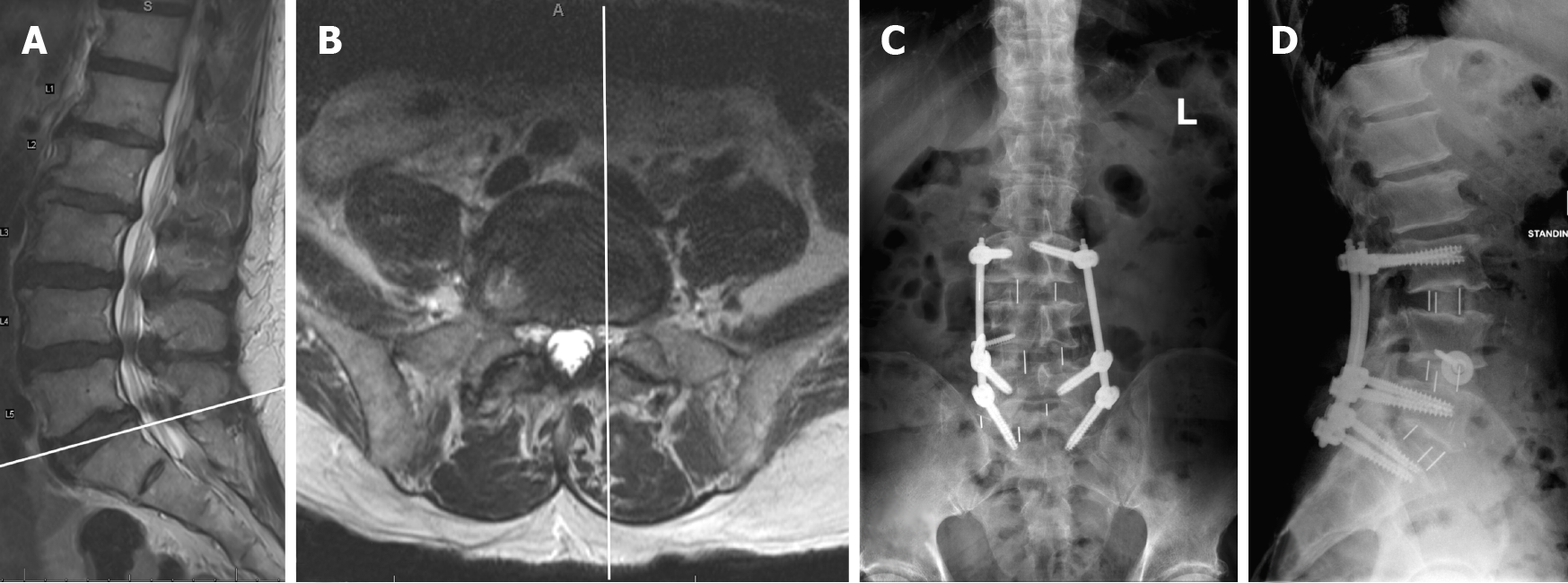
Figure 7 Preoperative magnetic resonance imaging sagittal (A) and axial (B) images and postoperative (C) anteroposterior and (D) lateral upright radiographs for Case 3.
Image A shows the scout line corresponding to the axial image B. The anteroposterior facet line through the medial border of the left L5-S1 facet joint runs lateral to the medial border of the left common iliac vein (CIV) (B). The fat plane is better visualized under the right CIV than under the left CIV (B). CIV: Common iliac vein.
- Citation: Berry CA. Nuances of oblique lumbar interbody fusion at L5-S1: Three case reports. World J Orthop 2021; 12(6): 445-455
- URL: https://www.wjgnet.com/2218-5836/full/v12/i6/445.htm
- DOI: https://dx.doi.org/10.5312/wjo.v12.i6.445


