Copyright
©The Author(s) 2021.
World J Orthop. Jun 18, 2021; 12(6): 445-455
Published online Jun 18, 2021. doi: 10.5312/wjo.v12.i6.445
Published online Jun 18, 2021. doi: 10.5312/wjo.v12.i6.445
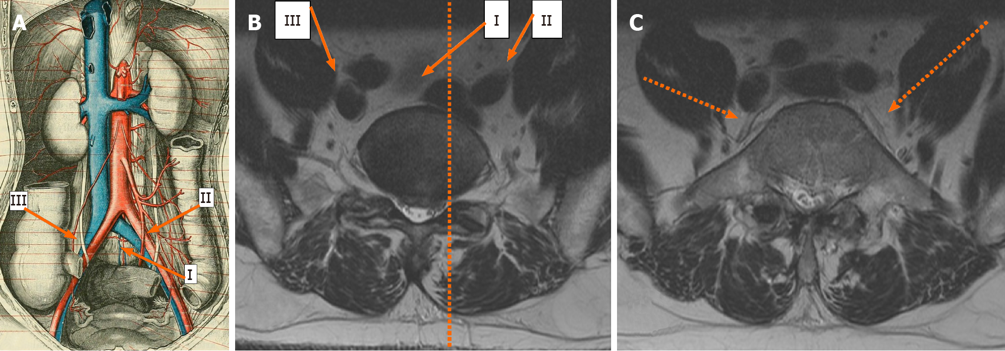
Figure 1 Vascular anatomy as depicted through an illustration showing the frontal aspect (A), and that depicted through magnetic resonance imaging axial sections (B, C).
Image B shows magnetic resonance imaging (MRI) axial section through the L5-S1 disc, and the facet line (dotted line) running anteroposteriorly through the medial border of the left L5-S1 facet joint cutting through the left common iliac vein. The three oblique approaches to L5-S1; namely the left intra-bifurcation (i), the left pre-psoas (ii), and the right pre-psoas (iii) are shown in A and B. Image C shows an MRI axial section through mid-L5 vertebral body and shows the left and right ilio-lumbar veins (dotted arrows).
- Citation: Berry CA. Nuances of oblique lumbar interbody fusion at L5-S1: Three case reports. World J Orthop 2021; 12(6): 445-455
- URL: https://www.wjgnet.com/2218-5836/full/v12/i6/445.htm
- DOI: https://dx.doi.org/10.5312/wjo.v12.i6.445


