Copyright
©The Author(s) 2021.
World J Orthop. May 18, 2021; 12(5): 338-345
Published online May 18, 2021. doi: 10.5312/wjo.v12.i5.338
Published online May 18, 2021. doi: 10.5312/wjo.v12.i5.338
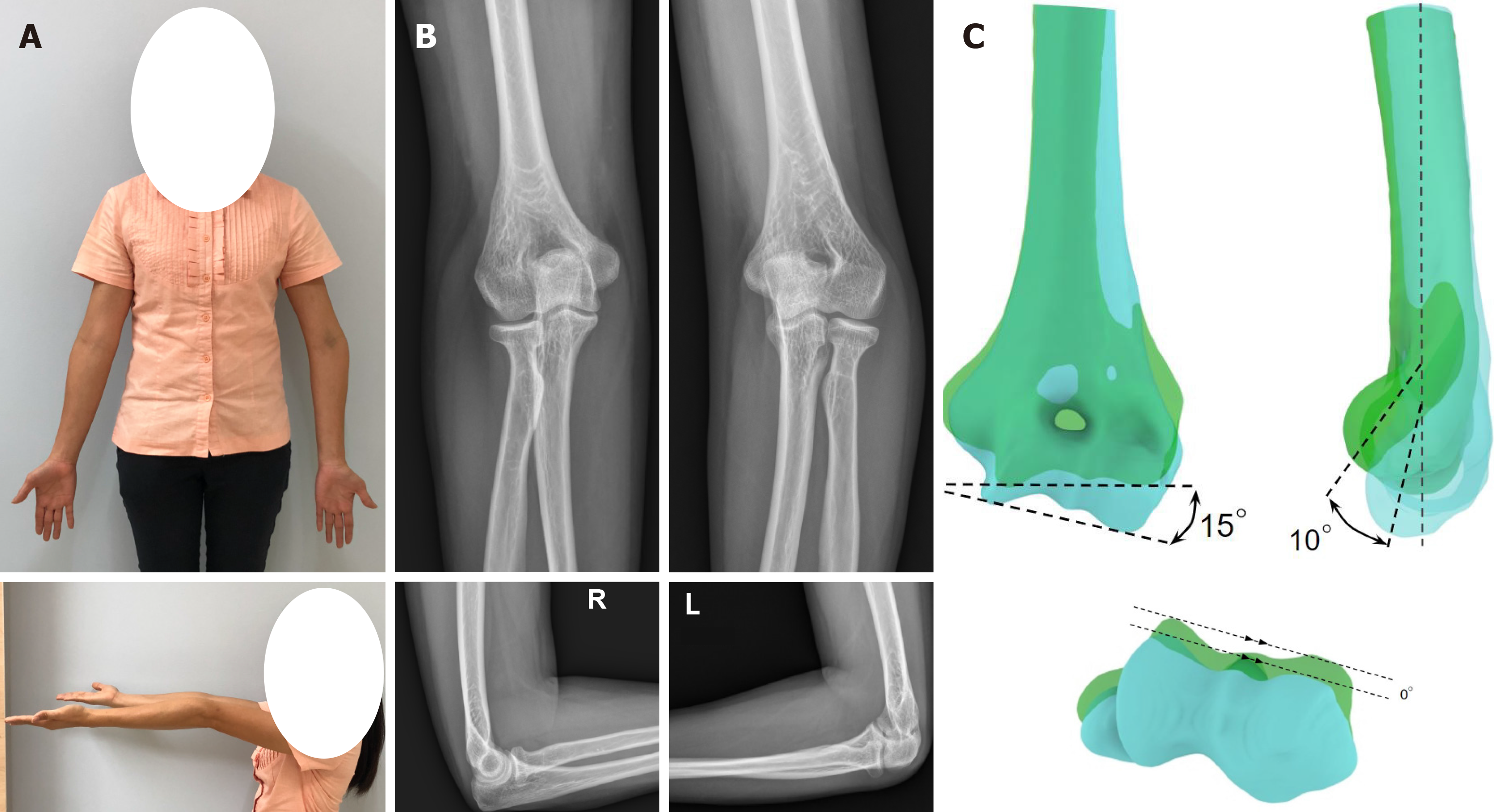
Figure 1 Preoperative evaluation.
A: Clinical pictures showing the cubitus varus and hypertension deformity of the left elbow; B: Preoperative radiographs in antero-posterior and lateral views showing a humerus-elbow-wrist angle of 15º varus and a shaft condylar angles of 35º on the left side, and those of 13º valgus and 45º, respectively; C: Three-dimensional computed tomography analysis with overlapping of the mirror image model from the right humerus (green color) and the deformed model from the left humerus (blue color). Anterior view showing a 15º-degree varus deformity. Lateral view showing a 10º-degree hyperextension deformity. Axial view showing no rotational deformity.
- Citation: Sri-utenchai N, Pengrung N, Srikong K, Puncreobutr C, Lohwongwatana B, Sa-ngasoongsong P. Three-dimensional printing technology for patient-matched instrument in treatment of cubitus varus deformity: A case report. World J Orthop 2021; 12(5): 338-345
- URL: https://www.wjgnet.com/2218-5836/full/v12/i5/338.htm
- DOI: https://dx.doi.org/10.5312/wjo.v12.i5.338


