Copyright
©The Author(s) 2021.
World J Orthop. May 18, 2021; 12(5): 270-291
Published online May 18, 2021. doi: 10.5312/wjo.v12.i5.270
Published online May 18, 2021. doi: 10.5312/wjo.v12.i5.270
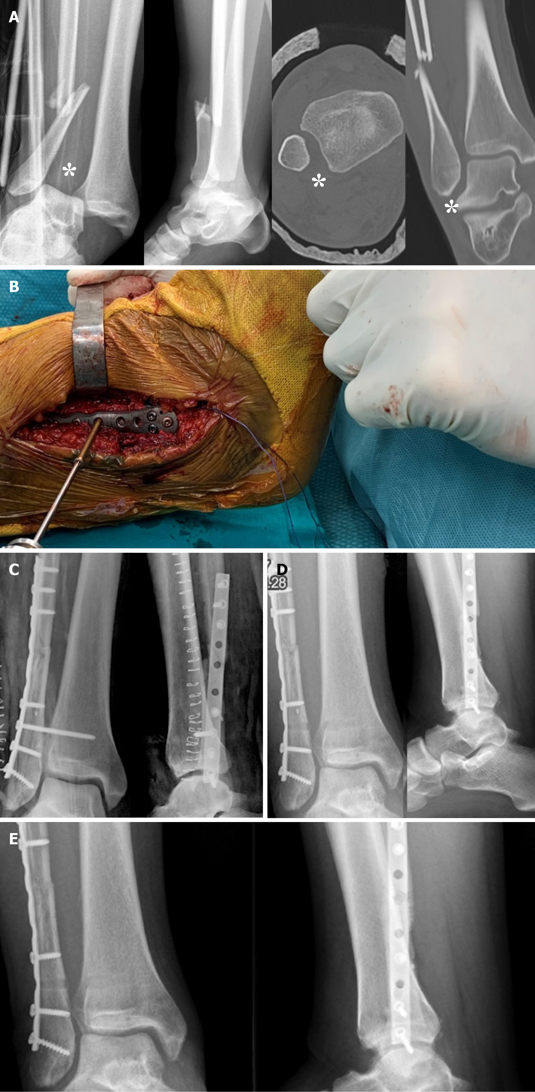
Figure 11 Type C ankle fracture.
A: Preoperative X-rays and computed tomography performed after closed reduction with diastasis (asterisks); B: Intraoperative positioning of the 3.5 mm intersyndesmotic screw with the foot positioned at 90° of dorsiflexion; C: Postoperative radiographs; D: Removal of the screw 2 mo after surgery; E: Views 6 mo after trauma with fracture consolidated.
- Citation: Pogliacomi F, De Filippo M, Casalini D, Longhi A, Tacci F, Perotta R, Pagnini F, Tocco S, Ceccarelli F. Acute syndesmotic injuries in ankle fractures: From diagnosis to treatment and current concepts. World J Orthop 2021; 12(5): 270-291
- URL: https://www.wjgnet.com/2218-5836/full/v12/i5/270.htm
- DOI: https://dx.doi.org/10.5312/wjo.v12.i5.270


