Copyright
©The Author(s) 2021.
World J Orthop. May 18, 2021; 12(5): 270-291
Published online May 18, 2021. doi: 10.5312/wjo.v12.i5.270
Published online May 18, 2021. doi: 10.5312/wjo.v12.i5.270
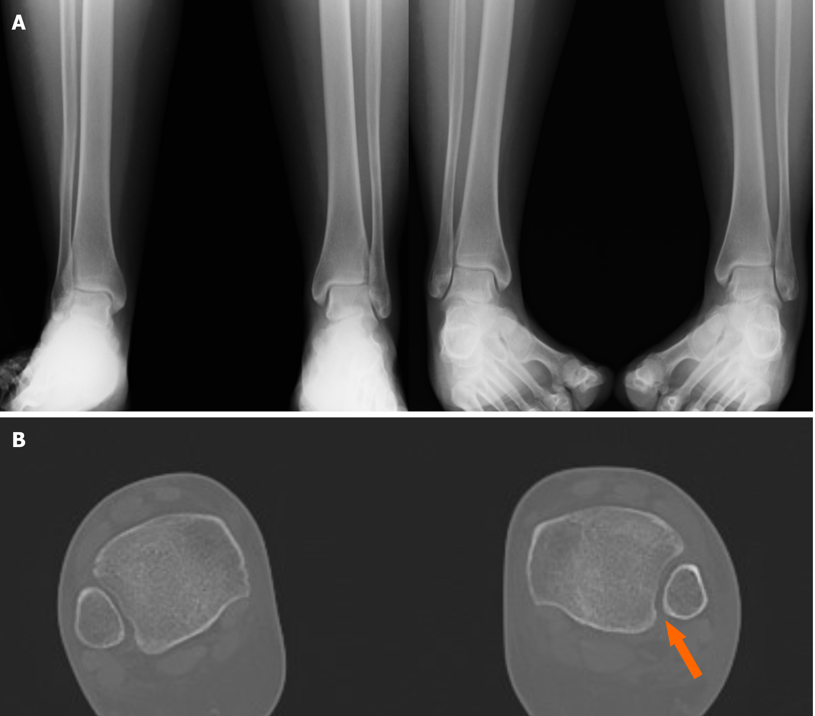
Figure 5 Suspect of a left syndesmotic lesion.
A: Comparative X-ray and based on the measurements of the tibiofibular clear space, tibiofibular overlap and medial clear space, the exam was considered negative; B: Computed tomography assessment showed a difference (> 2 mm) between both sides (arrow), demonstrating the presence of a syndesmotic lesion.
- Citation: Pogliacomi F, De Filippo M, Casalini D, Longhi A, Tacci F, Perotta R, Pagnini F, Tocco S, Ceccarelli F. Acute syndesmotic injuries in ankle fractures: From diagnosis to treatment and current concepts. World J Orthop 2021; 12(5): 270-291
- URL: https://www.wjgnet.com/2218-5836/full/v12/i5/270.htm
- DOI: https://dx.doi.org/10.5312/wjo.v12.i5.270


