Copyright
©The Author(s) 2021.
World J Orthop. Dec 18, 2021; 12(12): 970-982
Published online Dec 18, 2021. doi: 10.5312/wjo.v12.i12.970
Published online Dec 18, 2021. doi: 10.5312/wjo.v12.i12.970
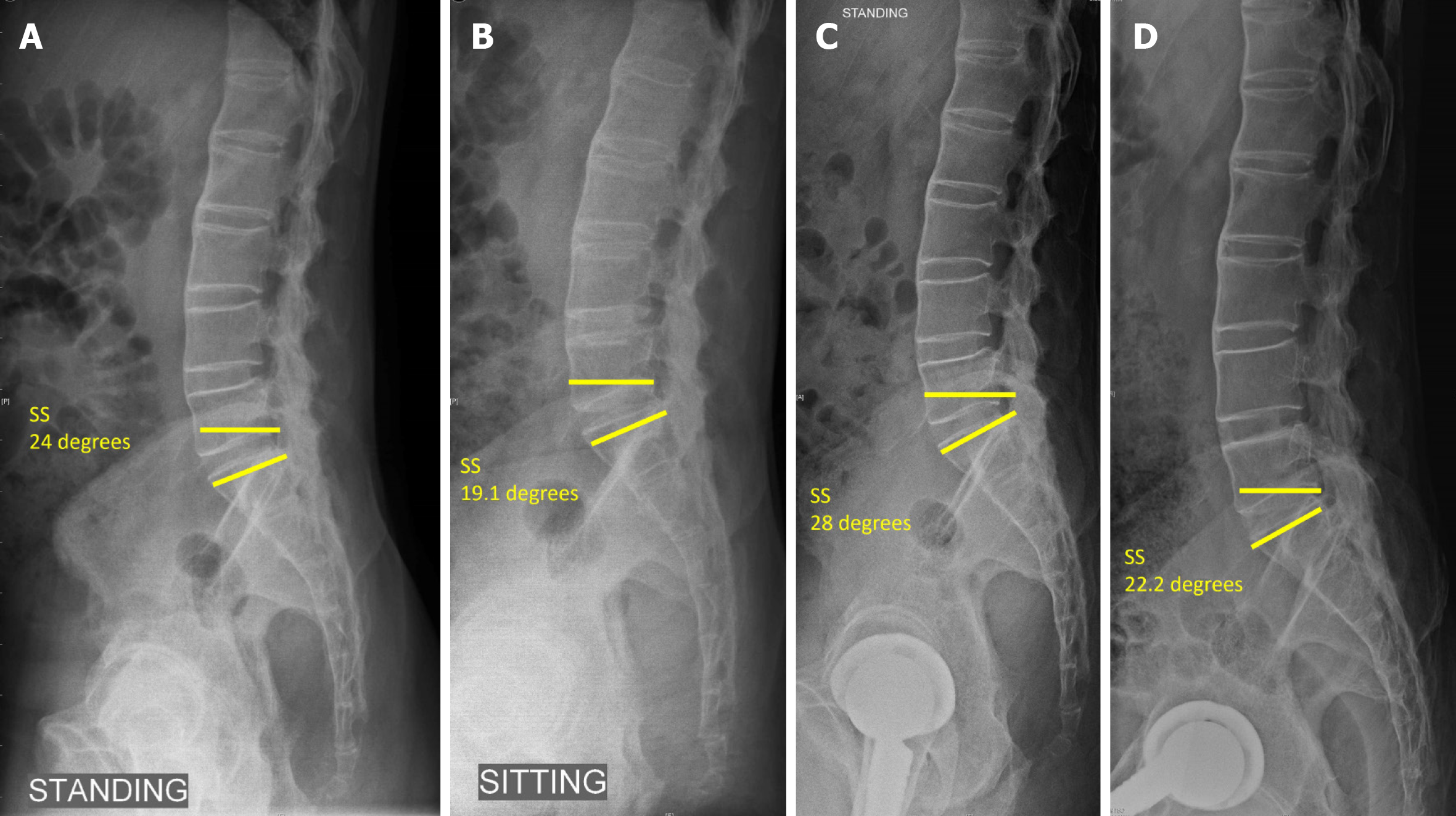
Figure 5 Lateral lumbosacral spine radiographs.
A: Pre op standing; B: Sitting compared to; C: Post op standing; D: Sitting showing the change in sacral slope < 10 degrees with reduced sacral slope indicating posterior pelvic tilt and stuck sitting pattern in ankylosing spondylitis (sacral slope < 30 degrees on sitting and standing typical of stuck sitting pattern). SS: Sacral slope.
- Citation: Oommen AT, Hariharan TD, Chandy VJ, Poonnoose PM, A AS, Kuruvilla RS, Timothy J. Total hip arthroplasty in fused hips with spine stiffness in ankylosing spondylitis. World J Orthop 2021; 12(12): 970-982
- URL: https://www.wjgnet.com/2218-5836/full/v12/i12/970.htm
- DOI: https://dx.doi.org/10.5312/wjo.v12.i12.970


