Copyright
©The Author(s) 2021.
World J Orthop. Dec 18, 2021; 12(12): 961-969
Published online Dec 18, 2021. doi: 10.5312/wjo.v12.i12.961
Published online Dec 18, 2021. doi: 10.5312/wjo.v12.i12.961
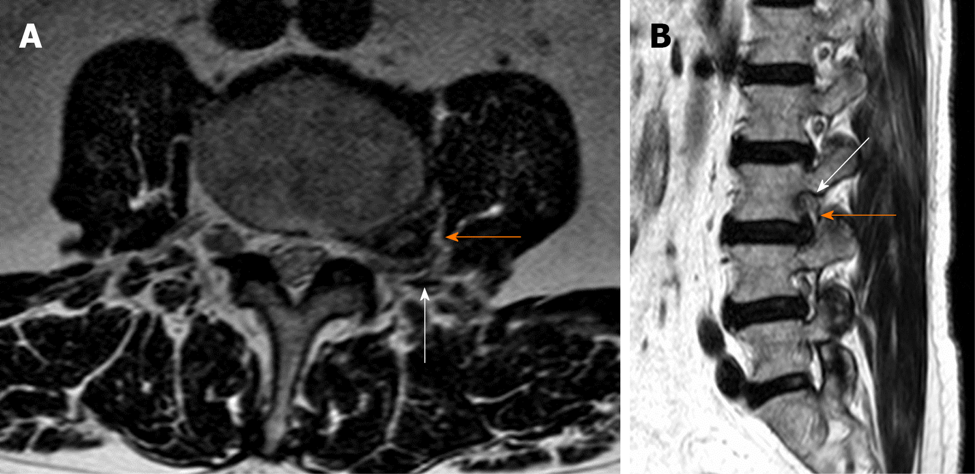
Figure 8 Axial (A) and sagittal (B) T2 magnetic resonance imaging showing a left L3-L4 extraforaminal far lateral lumbar disc herniations (orange arrow).
The L3 root is severely compressed against the posterior border of the neural foramen (white arrow).
- Citation: Berra LV, Di Rita A, Longhitano F, Mailland E, Reganati P, Frati A, Santoro A. Far lateral lumbar disc herniation part 1: Imaging, neurophysiology and clinical features. World J Orthop 2021; 12(12): 961-969
- URL: https://www.wjgnet.com/2218-5836/full/v12/i12/961.htm
- DOI: https://dx.doi.org/10.5312/wjo.v12.i12.961


