Copyright
©The Author(s) 2021.
World J Orthop. Nov 18, 2021; 12(11): 867-876
Published online Nov 18, 2021. doi: 10.5312/wjo.v12.i11.867
Published online Nov 18, 2021. doi: 10.5312/wjo.v12.i11.867
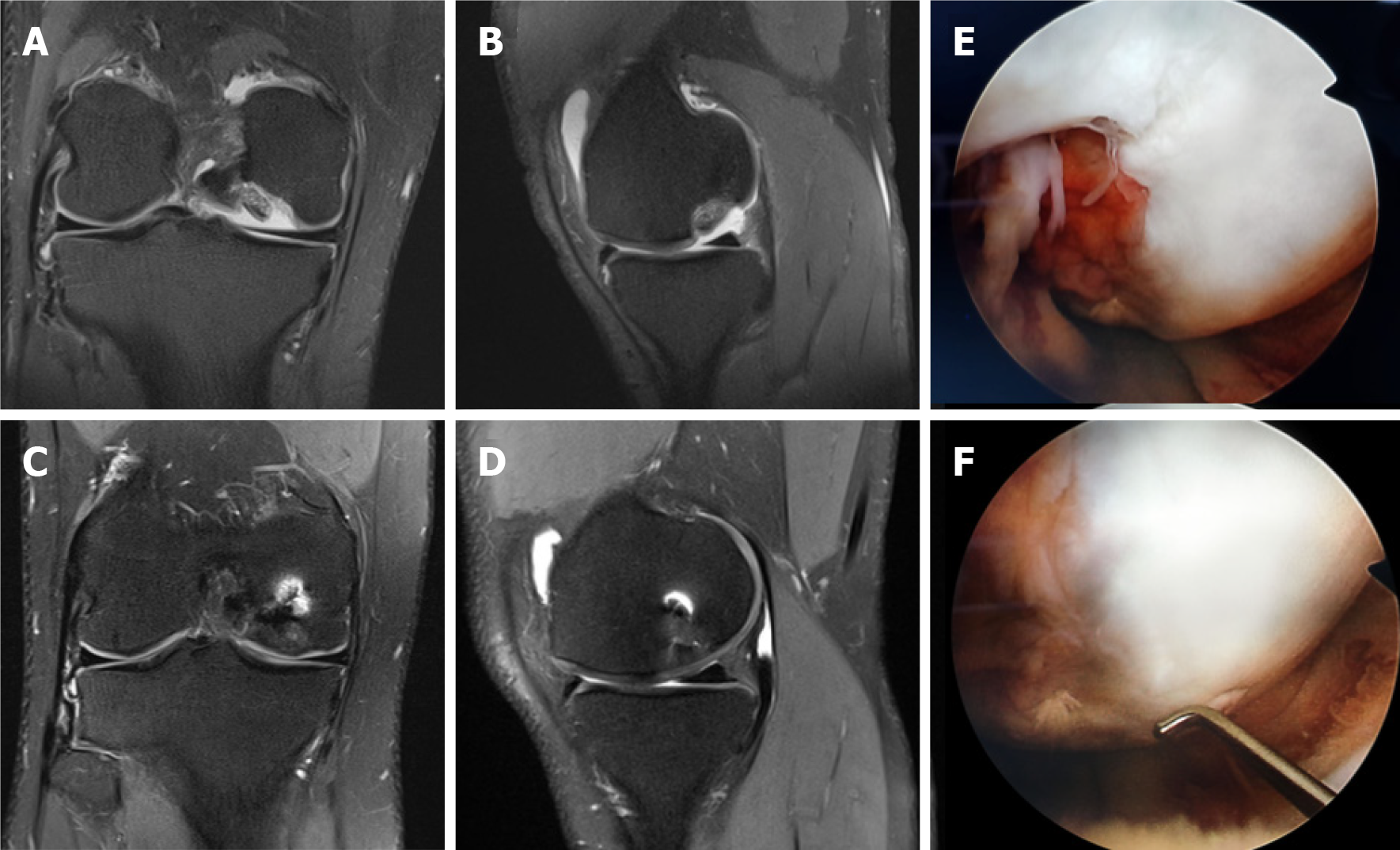
Figure 4 Magnetic resonance images and intraoperative images of a patient treated with the Autologous Tendon Transplantation technique.
A, B: Coronal and Sagittal T2 section magnetic resonance images (MRI) of a grade 4 osteochondral lesion involving articular surface of medial femoral condyle (3.4 cm2 size and 0.5 cm depth) in 24-year old male; C, D: 1-year follow-up coronal and sagittal T2 section MRI showing complete filling of the defect and establishment of smooth articular surface; E, F: Second look arthroscopy view at 1-year follow-up of grade 4 osteochondral lesion of medial femoral condyle showing filling of the defect with a well-integrated, smooth surfaced and stable regenerated cartilage.
- Citation: Turhan AU, Açıl S, Gül O, Öner K, Okutan AE, Ayas MS. Treatment of knee osteochondritis dissecans with autologous tendon transplantation: Clinical and radiological results. World J Orthop 2021; 12(11): 867-876
- URL: https://www.wjgnet.com/2218-5836/full/v12/i11/867.htm
- DOI: https://dx.doi.org/10.5312/wjo.v12.i11.867


