Copyright
©The Author(s) 2020.
World J Orthop. Feb 18, 2020; 11(2): 137-144
Published online Feb 18, 2020. doi: 10.5312/wjo.v11.i2.137
Published online Feb 18, 2020. doi: 10.5312/wjo.v11.i2.137
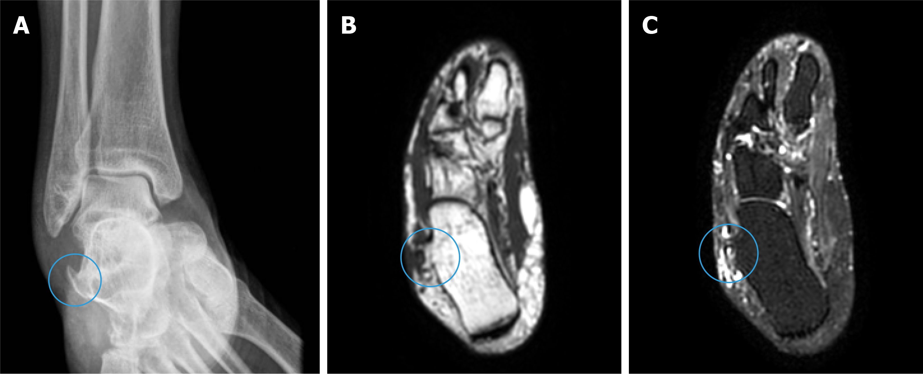
Figure 2 Preoperative plain radiograph and magnetic resonance imaging evaluation for surgical planning.
A: Ankle plain radiograph anteroposterior view showing a sharp hypertrophic peroneal tubercle (blue circle); B and C: Ankle magnetic resonance imaging T1- and T2-weighted axial images (B and C respectively) demonstrated the hypertrophic peroneal tubercle with underlying bone edema (blue circles).
- Citation: Nishikawa DRC, Duarte FA, Saito GH, de Cesar Netto C, Fonseca FCP, Miranda BR, Monteiro AC, Prado MP. Minimally invasive tenodesis for peroneus longus tendon rupture: A case report and review of literature. World J Orthop 2020; 11(2): 137-144
- URL: https://www.wjgnet.com/2218-5836/full/v11/i2/137.htm
- DOI: https://dx.doi.org/10.5312/wjo.v11.i2.137


